. 2015 Feb;22(2):158-66.
doi: 10.1038/nsmb.2945. Epub 2015 Jan 12.

Tetrameric Ctp1 coordinates DNA binding and DNA bridging in DNA double-strand-break repair

Affiliations

Tetrameric Ctp1 coordinates DNA binding and DNA bridging in DNA double-strand-break repair

Sara N Andres et al. Nat Struct Mol Biol. 2015 Feb.

Abstract

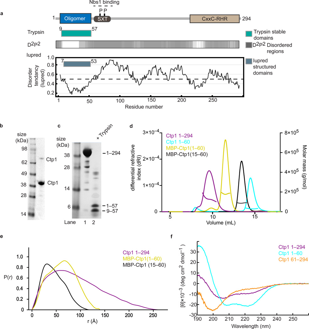
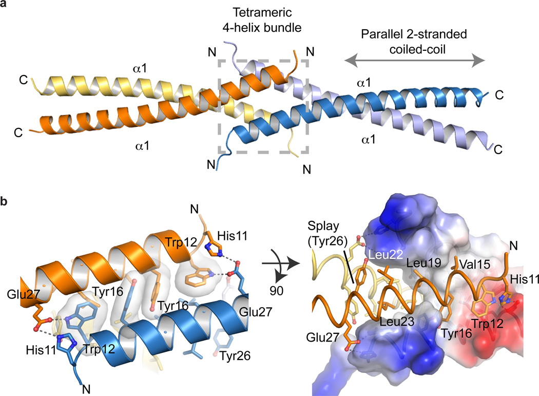
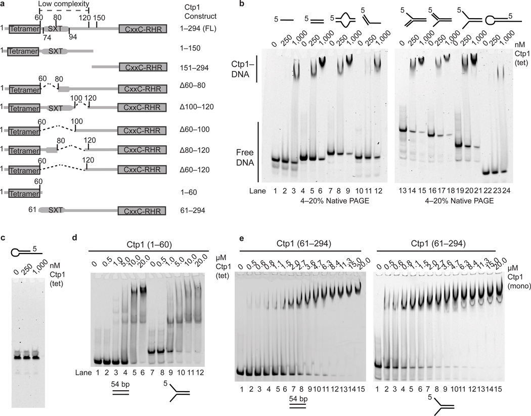
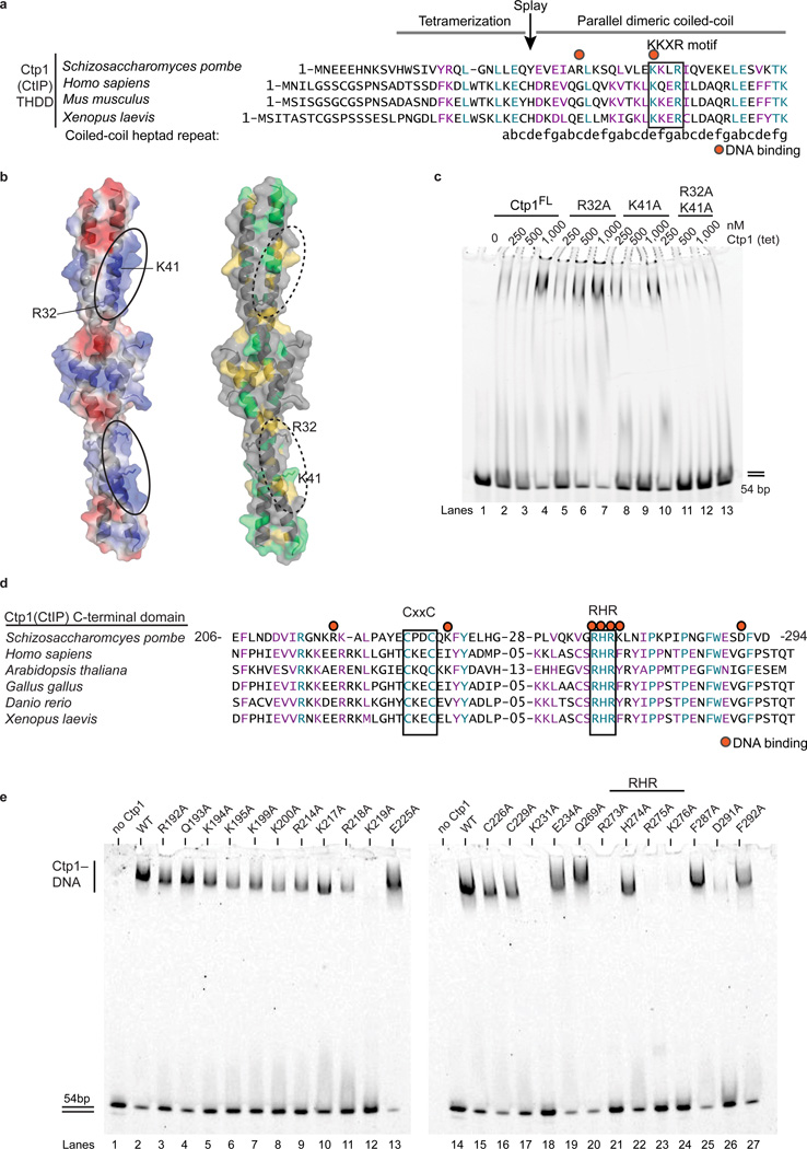
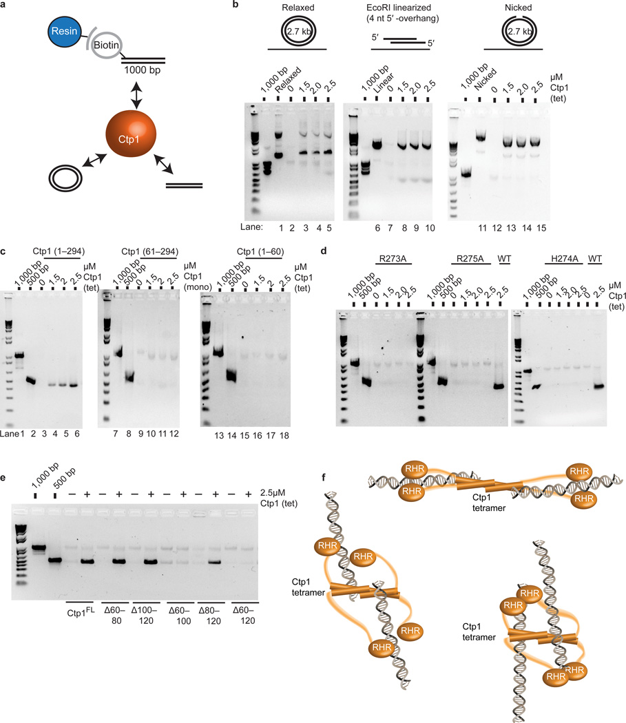
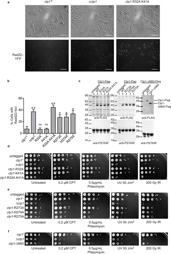
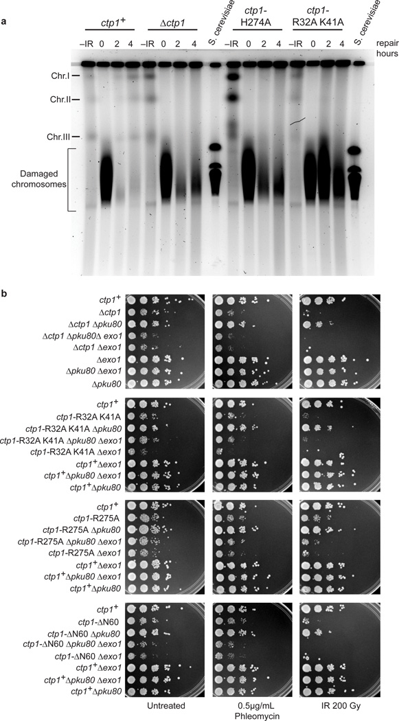
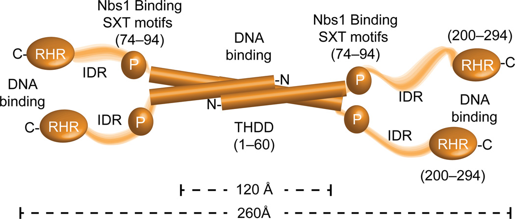
Ctp1 (also known as CtIP or Sae2) collaborates with Mre11-Rad50-Nbs1 to initiate repair of DNA double-strand breaks (DSBs), but its functions remain enigmatic. We report that tetrameric Schizosaccharomyces pombe Ctp1 contains multivalent DNA-binding and DNA-bridging activities. Through structural and biophysical analyses of the Ctp1 tetramer, we define the salient features of Ctp1 architecture: an N-terminal interlocking tetrameric helical dimer-of-dimers (THDD) domain and a central intrinsically disordered region (IDR) linked to C-terminal 'RHR' DNA-interaction motifs. The THDD, IDR and RHR are required for Ctp1 DNA-bridging activity in vitro, and both the THDD and RHR are required for efficient DSB repair in S. pombe. Our results establish non-nucleolytic roles of Ctp1 in binding and coordination of DSB-repair intermediates and suggest that ablation of human CtIP DNA binding by truncating mutations underlie the CtIP-linked Seckel and Jawad syndromes.


Figures

Figure 1
Figure 1
Tetrameric Ctp1 is an intrinsically disordered protein (a) Domain structure of Ctp1 with proteolytic mapping and order or disorder predictions. (b) Purified full-length recombinant Ctp1. Monomeric and dimeric Ctp1 species are labeled. (c) Limited trypsin proteolysis of Ctp1. Trypsinolysis produced two stable fragments that mass spectrometry identified as Ctp11–57 and Ctp19–57 (d) SEC-MALS traces of differential refractive index and molar mass for Ctp1, Ctp11–60, N-terminal maltose-binding protein (MBP)-tagged Ctp11–60 and N-terminal MBP-tagged Ctp115–60 (e) Ctp1 SAXS pair distribution functions of Ctp1, MBP-tagged Ctp11–60 and Ctp115–60 (f) Circular dichroism spectra of Ctp1, Ctp1 1–60 and Ctp161–294 . θ, mean residue ellipticity. Original gels can be found in Supplementary Data Set 1.
Figure 2
Figure 2
X-ray crystal structure of the Ctp1 N-terminal tetramerization domain (a) Cartoon representation of Ctp1 highlighting the tetrameric 4-helix bundle (gray dotted-line box) and the parallel 2-stranded coiled-coil (gray arrow). (b) The Ctp1 aromatic hooks. His11, Trp12 and Tyr16 interact with opposing helices to stabilize the tetrameric interface. The electrostatic surface representation of Ctp1 illustrates the transition from tetrameric 4-helix bundle to dimeric coiled-coil by the Tyr26 splay. Blue, electropositive surface; red, electronegative surface; white, hydrophobic.
Figure 3
Figure 3
Ctp1 binds DNA. (a) Domain organization and schematic of Ctp1 N-terminal, C-terminal and internal deletion constructs. (b) Ctp1 binds assorted DNA substrates. (c) Ctp1 displays no appreciable nuclease activity on a DNA hairpin substrate. (d) Ctp11–60 binding double-stranded and forked DNA. (e) Ctp161–294 binds double-stranded and forked DNA. Ctp1 concentrations expressed in terms of molarity of the Ctp1 tetramer (tet) or monomer (mono) as labeled. Experiments were repeated 3 times for (b), (d), and (e), and 2 times for (c). Original gels can be found in Supplementary Data Set 1.
Figure 4
Figure 4
Identifying Ctp1–DNA interaction motifs. (a) Structure-based sequence alignment of the Ctp1 THDD domain. Conserved identical residues are highlighted blue, and similar residues are highlighted purple. Red dots indicate residues targeted for mutagenesis, and the black box outlines the conserved KKxR motif. The position of the Ctp1 coiled-coil “a”→“g” heptad repeat is indicated below the sequence. (b) Electrostatic surface potential (left) and conserved surface mapping (right) of the putative Ctp1 THDD DNA binding interface. Red, electropositive; blue, electronegative; green, conserved identical residues; yellow, conserved similar residues. (c) Ctp1 THDD mutants binding DNA. Ctp1 concentrations expressed in terms of molarity of the Ctp1 tetramer (tet). (d) Sequence alignment of the Ctp1 C-terminal domain. Same coloring as in (a). Red dots designate DNA-binding amino acids. Black boxes outline conserved CxxC and RHR motifs. (e) Alanine scanning mutagenesis to probe DNA binding amino acids in the Ctp1 C-terminal region. Experiments were repeated 3 times for (c), (e). Original gels can be found in Supplementary Data Set 1.
Figure 5
Figure 5
Ctp1 bridges DNA (a) Bridging assay schematic. Ctp1 binds a biotinylated 1000 bp DNA substrate linked to magnetic beads and bridges to various substrates. (b) Bridging activity of Ctp1 to plasmid DNA substrates. “1000 bp” denotes biotinylated substrate prior to incubation with protein and DNA bridging targets. (c) DNA bridging activity of Ctp1, Ctp161–294 and Ctp11–60 to a 500bp dsDNA. (d) Bridging activity of Ctp1 C-terminal RHR mutants. WT, wildtype. (e) Bridging activity of Ctp1 internal deletions of the IDR region. (f) Model of Ctp1 binding and bridging a DNA DSB. Three possible Ctp1 DNA bridging architectures are diagrammed. Ctp1 concentrations expressed in terms of molarity of the Ctp1 tetramer (tet) or monomer (mono) as labeled. Experiments were repeated 3 times for (b), (c), (d), and (e), and twice for: EcoRI-linearized in (b), H274A in (d) and Ctp1Δ60–80 in (e). Original gels can be found in Supplementary Data Set 1.
Figure 6
Figure 6
Sensitivity of Ctp1 THDD and RHR mutations to DNA damaging agents (a) Representative images from n=3 cell culture replicates of Rad22-YFP foci in ctp1+, ctp1Ä, and ctp1-R32A K41A strains. Scale bars are 20 µm. (b) Levels of spontaneous Rad22-YFP foci in S. pombe harboring THDD and RHR mutations. Mean values shown. Error bars, s.d. (n=3 cell cultures) * P-value < 0.01; ** P-value < 0.001 compared to ctp1+ by two-tailed Student’s t test. ns, not significant. (c) Immunoblot (anti-Flag) of Ctp1-Flag tagged strains used in panels (a), (b), (d)(f). Anti-PSTAIR is the loading control. (d) Sensitivity of Ctp1 N-terminal DNA binding mutants to DNA damaging agents. Representative images from n=3 technical replicates. (e) Sensitivity of Ctp1 C-terminal RHR mutants to DNA damaging agents. Images are representative of n=3 technical replicates . (f) Ctp1 N-terminal 60 amino acid deletion mutant sensitivity to DNA damaging agents . Representative images from n=3 technical replicates. Original blots can be found in Supplementary Data Set 1.
Figure 7
Figure 7
Ctp1 THDD and RHR mutations effects on S. pombe DSB repair. (a) Damage and repair of IR damaged S. pombe chromosomes. –IR, chromosomes prior to treatment with IR; S. cerevisiae chromosomes were used as size marker. Image is representative of n=2 cell culture replicates. (b) Suppression of Ctp1 RHR, THDD, and N-terminal deletion mutants by deleting Ku80, and rescue by Exo1. Representative images from n=3 technical replicates. Original full-size gel shown.
Figure 8
Figure 8
Overall model for Ctp1 molecular architecture from X-ray structures, SAXS and biophysical analysis.

References

    1. Williams RS, Williams JS, Tainer JA. Mre11-Rad50-Nbs1 is a keystone complex connecting DNA repair machinery, double-strand break signaling, and the chromatin template. Biochem. Cell Biol. 2007;85:509–520. - PubMed
    1. Pommier Y. Drugging topoisomerases: lessons and challenges. ACS Chem. Biol. 2013;8:82–95. - PMC - PubMed
    1. You Z, et al. CtIP links DNA double-strand break sensing to resection. Mol. Cell. 2009;36:954–969. - PMC - PubMed
    1. Williams RS, et al. Nbs1 flexibly tethers Ctp1 and Mre11-Rad50 to coordinate DNA double-strand break processing and repair. Cell. 2009;139:87–99. - PMC - PubMed
    1. Limbo O, et al. Ctp1 is a cell-cycle-regulated protein that functions with Mre11 complex to control double-strand break repair by homologous recombination. Mol. Cell. 2007;28:134–146. - PMC - PubMed

Online Methods References

    1. Stols L, et al. A new vector for high-throughput, ligation-independent cloning encoding a tobacco etch virus protease cleavage site. Protein Expr. Purif. 2002;25:8–15. - PubMed
    1. Sreerama N, Woody RW. A self-consistent method for the analysis of protein secondary structure from circular dichroism. Anal. Biochem. 1993;209:32–44. - PubMed
    1. Sreerama N, Venyaminov SY, Woody RW. Estimation of the number of alpha-helical and beta-strand segments in proteins using circular dichroism spectroscopy. Protein Sci. 1999;8:370–380. - PMC - PubMed
    1. Whitmore L, Wallace BA. DICHROWEB, an online server for protein secondary structure analyses from circular dichroism spectroscopic data. Nucleic Acids Res. 2004;32:W668–W673. - PMC - PubMed
    1. Otwinowski Z, Minor W. Processing of X-ray diffraction data collected in osciallation mode. In: Carter CW Jr, Sweet RM, editors. Methods in Enzymology. Vol. 276. Academic Press; 1997. pp. 307–326. - PubMed

Publication types

MeSH terms

Substances

Associated data