

Accelerated detection of viral particles by combining AC electric field effects and micro-Raman spectroscopy
- PMID: 25580902
- PMCID: PMC4327063
- DOI: 10.3390/s150101047
Accelerated detection of viral particles by combining AC electric field effects and micro-Raman spectroscopy
Abstract
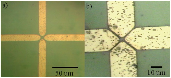
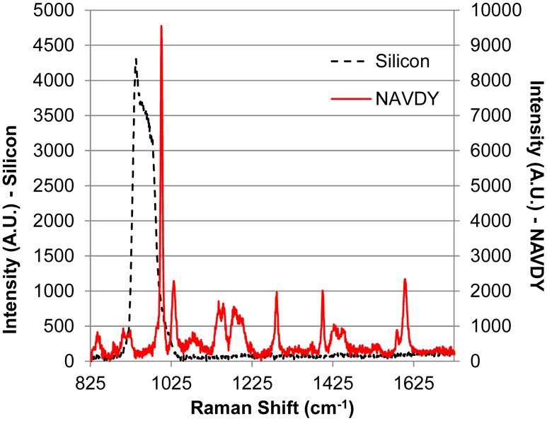
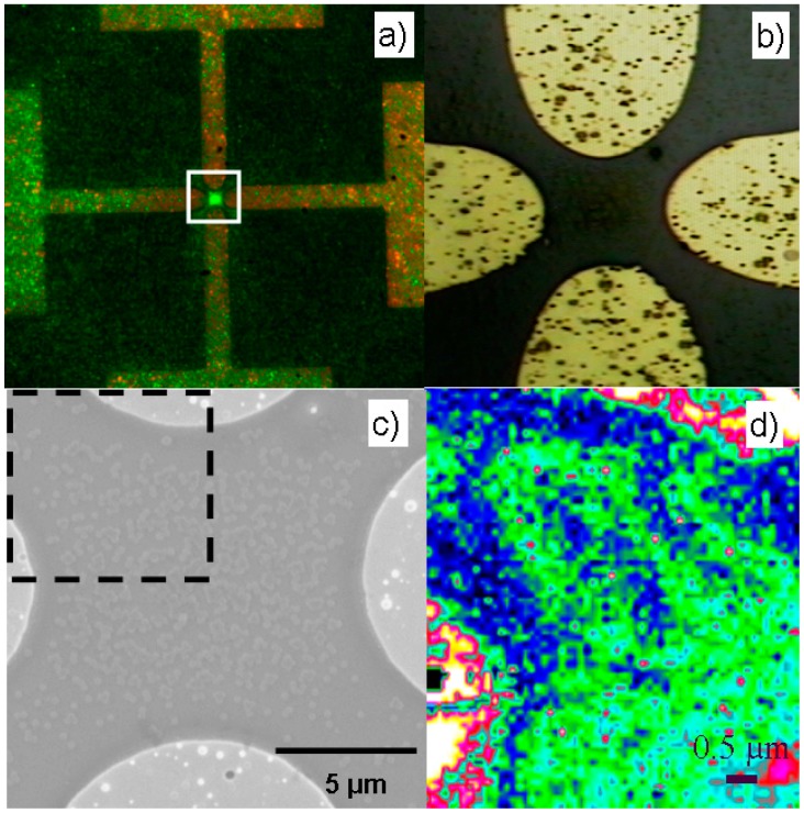
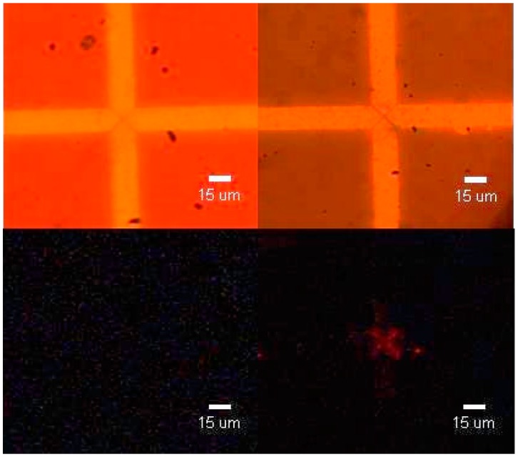
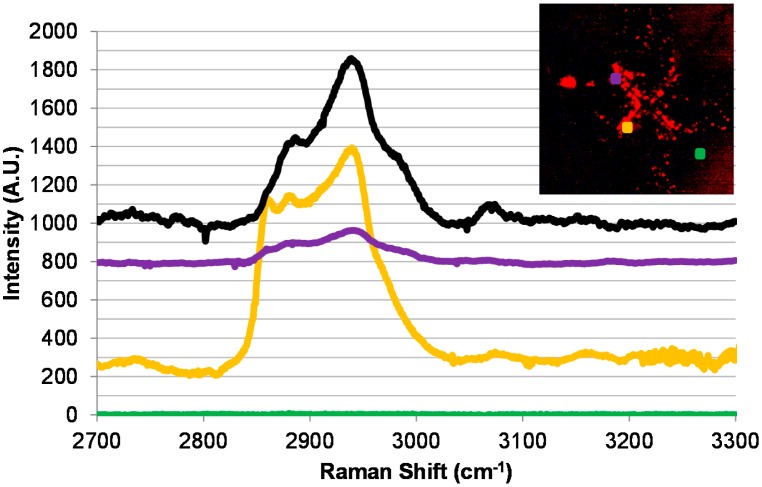
A detection method that combines electric field-assisted virus capture on antibody-decorated surfaces with the "fingerprinting" capabilities of micro-Raman spectroscopy is demonstrated for the case of M13 virus in water. The proof-of-principle surface mapping of model bioparticles (protein coated polystyrene spheres) captured by an AC electric field between planar microelectrodes is presented with a methodology for analyzing the resulting spectra by comparing relative peak intensities. The same principle is applied to dielectrophoretically captured M13 phage particles whose presence is indirectly confirmed with micro-Raman spectroscopy using NeutrAvidin-Cy3 as a labeling molecule. It is concluded that the combination of electrokinetically driven virus sampling and micro-Raman based signal transduction provides a promising approach for time-efficient and in situ detection of viruses.
Figures
References
-
- Yang L., Bashir R. Electrical/Electrochemical Impedance for the Rapid Detection of Foodborne Pathogenic Bacteria. Biotechnol. Adv. 2008;26:135–150. - PubMed
-
- Docoslis A., Espinoza L.A.T., Zhang B., Cheng L., Israel B.A., Alexandridis P., Abbott N.L. Using Nonuniform Electric Fields to Accelerate the Transport of Viruses to Surfaces from Media of Physiological Ionic Strength. Langmuir. 2007;23:3840–3848. - PubMed
-
- Morgan H., Green N.G. Dielectrophoretic Manipulation of Rod-Shaped Viral Particles. J. Electrost. 1997;42:279–293.
-
- Roy R., Tomkins M., Docoslis A. Enhancing the Performance of Surface-Based Biosensors by AC Electrokinetic Effects—A Review. In: Serra P.A., editor. Biosensors—Emerging Materials and Applications. InTech; Rijeka, Croatia: 2011. pp. 243–264.
-
- Morgan H., Green N.G. AC Electrokinetics: Colloids and Nanoparticles. 2nd ed. Research Studies Press Ltd; Baldock, UK: 2003.
Publication types
MeSH terms
Substances
LinkOut - more resources
Full Text Sources
Other Literature Sources
Molecular Biology Databases
