Review
. 2015:33:257-90.
doi: 10.1146/annurev-immunol-032414-112240. Epub 2015 Jan 2.

Innate immune pattern recognition: a cell biological perspective

Affiliations
Review

Innate immune pattern recognition: a cell biological perspective

Sky W Brubaker et al. Annu Rev Immunol. 2015.

Abstract

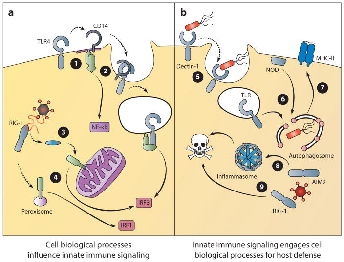
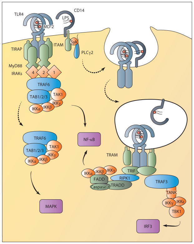
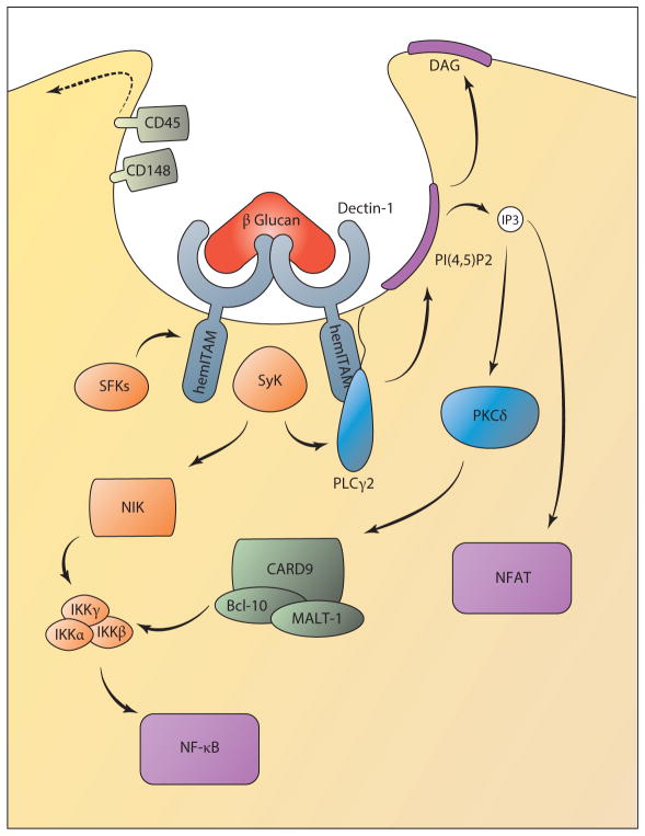
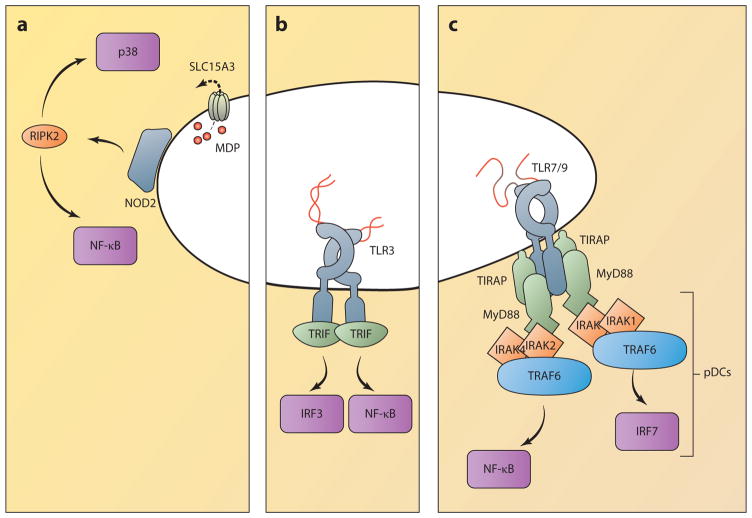
Receptors of the innate immune system detect conserved determinants of microbial and viral origin. Activation of these receptors initiates signaling events that culminate in an effective immune response. Recently, the view that innate immune signaling events rely on and operate within a complex cellular infrastructure has become an important framework for understanding the regulation of innate immunity. Compartmentalization within this infrastructure provides the cell with the ability to assign spatial information to microbial detection and regulate immune responses. Several cell biological processes play a role in the regulation of innate signaling responses; at the same time, innate signaling can engage cellular processes as a form of defense or to promote immunological memory. In this review, we highlight these aspects of cell biology in pattern-recognition receptor signaling by focusing on signals that originate from the cell surface, from endosomal compartments, and from within the cytosol.

Keywords: ALR; CLR; NLR; RLR; TLR.


Figures

Figure 1
Figure 1
Implications of cell biological processes on innate immune signaling. The trafficking of PRRs and the location of innate immune signaling can regulate signal transduction and dictate the outcome of microbial detection (a). ➊ TLR4 undergoes trafficking to specialized regions of the plasma membrane to activate MyD88-dependent signaling. ➋ CD14 mediates the endocytosis of TLR4, which results in TRIF-dependent signaling from the endosome. ➌ RIG-I undergoes trafficking to the site of MAVS signaling on mitochondria. ➍ MAVS signaling from mitochondria induces a type I and III IFN response, whereas signaling from peroxisomes induces the transcription of type III IFN only. Alternatively, innate immune signaling can engage cell biological processes to control infection (b). ➎ Dectin signaling can induce phagocytosis as an effective means of microbial clearance. ➏ NODs and TLRs recruit regulators of autophagy to the site of bacterial entry for pathogen elimination. ➐ Autophagosomes can deliver antigens of intracellular pathogens for MHC-II presentation to coordinate adaptive immune responses. ➑ Inflammasome activation induces a rapid form of cell death known as pyroptosis, which can limit the replication of intracellular pathogens. ➒ The RLR/MAVS signaling pathway can also induce cell death to limit viral replication. (Solid lines indicate signal transduction; dotted lines indicate trafficking events.) (Abbreviations: MAVS, mitochondrial antiviral signaling protein; MHC-II, major histocompatibility complex class II; NOD, nucleotide-binding oligomerization domain; RIG-I, retinoic acid–inducible gene I; TRIF, TIR domain–containing adaptor-inducing IFN-β.)
Figure 2
Figure 2
TLR4 signals from the plasma membrane and endosomes. TLR4 requires translocation to lipid rafts enriched with TIRAP for signaling from the plasma membrane. This facilitates interactions with MyD88 upon ligand binding for the formation of the myddosome containing MyD88, TIRAP, and IRAKs. The IRAKs recruit the E3 ubiquitin ligase TRAF6, which interacts with a complex formed by TAB1, TAB2, TAB3, and TAK1. This complex regulates NF-κB activation via IKKs. TAK1 release into the cytoplasm also directs MAPK activation. CD14 controls the movement of TLR4 from the plasma membrane into endosomes through the activation of ITAM, Syk, and PLCγ2. From endosomes, TLR4 interacts with the sorting adaptor TRAM and the signaling adaptor TRIF to sustain NF-κB activation and to induce IRF3-mediated type I IFN production. TRIF-dependent NF-κB activation may proceed via the proteins RIPK1, TRADD, and the caspase-8 complex. IRF3 activation controls type I IFN production and requires TRAF3 recruitment to TRIF. TRAF3 then interacts with TANK (or TANK-related proteins) to recruit IKKγ, IKKε, and TBK1, which activate IRF3. (Solid lines indicate signal transduction; dotted lines indicate trafficking events.) (Abbreviations: FADD, Fas-associated protein with death domain; IKK, IκB kinase; IRAK, interleukin-1 receptor-associated kinase; IRF, IFN regulatory factor; ITAM, immunoreceptor tyrosine-based activation motif; MAPK, mitogen-activated protein kinase; NF-κB, nuclear factor-κB; PLCγ2, phospholipase Cγ2; RIPK1, receptor-interacting serine/threonine-protein kinase 1; TAB, TAK1-binding protein; TAK, TGF-β-activated kinase; TANK, TRAF family member–associated NF-κB activator; TBK, TANK-binding kinase; TIRAP, TIR-containing adaptor protein; TRADD, TNF receptor type 1–associated death domain; TRAF, TNF receptor–associated factor.)
Figure 3
Figure 3
Dectin-1 signaling controls NF-κB and NFAT activation. Upon ligand binding, the hemITAM domain of Dectin-1 is phosphorylated by Src family kinases (SFKs), which recruit the signaling kinase Syk. This requires trafficking of the phosphatases CD45 and CD148 away from the forming phagosome. Syk activation can control PLCγ2 activity to induce the formation of IP3 and DAG. IP3-mediated Ca2+ release from the endoplasmic reticulum induces a CRAC-dependent Ca2+ influx and consequent NFAT activation. Ca2+ and DAG also regulate PKCδactivation, which controls canonical NF-κB activation following induction of a CARD9/Bcl-10/MALT-1 complex. In parallel, Syk also controls noncanonical NF-κB activation through NIK. (Solid lines indicate signal transduction; dotted lines indicate trafficking events.) (Abbreviations: CARD, caspase recruitment domain; CRAC, calcium-release-activated calcium; DAG, diacylglycerol; IKK, IκB kinase; IP3, inositol 1,4,5-trisphosphate; ITAM, immunoreceptor tyrosine-based activation motif; NFAT, nuclear factor of activated T cells; NIK, NF-κB-inducing kinase; PKC, protein kinase C.)
Figure 4
Figure 4
Innate immune signaling from endosomes. (a) NOD receptors associate with endosomal membranes, where they are positioned to encounter PAMPs, as microbes escape from endosomes or as they are pumped out by transporters such as SLC15A3. NOD signaling activates RIPK2, which triggers proinflammatory cytokine production through NF-κB and MAPK activation. (b) TLR3 responds to double-stranded RNA and triggers an enzymatic signaling cascade through the adaptor protein TRIF. Unlike TLR4, TLR3 does not require the adaptor TRAM, but it can similarly activate IRF3 to produce type I IFN and ISGs, as well as NF-κB, to produce proinflammatory cytokines. (c) Other endosomal TLRs principally activate NF-κB through the adaptors MyD88 and TIRAP. In pDCs, TLR7 and -9 trigger IFN and ISG production through the activation of IRF7. (Abbreviations: IFN, interferon; IRAK, interleukin-1 receptor-associated kinase; IRF, IFN regulatory factor; ISG, IFN-stimulated gene; MAPK, mitogen-activated protein kinase; MDP, muramyl dipeptide; NOD, nucleotide-binding oligomerization domain; PAMP, pathogen-associated molecular pattern; pDC, plasmacytoid dendritic cell; RIPK2, RIP2 kinase; TIRAP, TIR-containing adaptor protein; TRAF, TNF receptor–associated factor; TRIF, TIR domain–containing adaptor-inducing IFN-β.)
Figure 5
Figure 5
The biosynthetic pathway: nucleic acid detection from within the cytosol. (a) The RLRs detect pathogen-derived RNA within the cytosol to induce the production of IFN and proinflammatory cytokines. The TBK1, IKK, and MAVS pathways lead to activation of the transcription factors for the induction of IFN and other cytokine genes. ➊ RIG-I signal transduction is regulated by TRIM25- and RIPLET-mediated ubiquitination and translocation to the site of signaling by 14-3-3ε. ➋ MAVS activity is regulated by polymerization, and signaling from mitochondria results in production of type I and III IFN, whereas peroxisomal signaling induces the production only of type III IFN. (b) cGAS and the ALRs detect pathogen-derived DNA from within the cytosol and nucleus to induce the production of IFN. ➌ In the presence of DNA, the enzyme cGAS converts ATP and GTP to the cyclic dinucleotide cGAMP. ➍ Production of cGAMP induces the activation and trafficking of STING to poorly defined sites of signaling in an ATG9- and VSP34-dependent manner. TBK1 is recruited to this site of signaling to induce the production of type I IFN. ➎ Viral DNA within the nucleus can be detected by IFI16 for the production of type I IFN in a STING-dependent manner. Therefore, the trafficking of this receptor or another factor from the nucleus to the cytosol may regulate IFI16-dependent signaling. (c) The inflammasome-mediated response to DNA within the cytosol. ➏ AIM2 detection of cytosolic DNA activates inflammasome formation, which induces cell death and the maturation of IL-1β. ➐ Secretion of this cytokine requires a noncanonical route that is independent of trafficking through the endoplasmic reticulum and Golgi apparatus. Several possibilities have been proposed for IL-1β release that include routes through lysosomal compartments, multivesicular bodies, or pores created during pyroptotic cell death. (Solid lines indicate signal transduction; dotted lines indicate trafficking events.) (Abbreviations: AIM, absent in melanoma; cGAMP, cyclic di-GMP/AMP; cGAS, cyclic GMP-AMP synthase; IFI, interferon, γ-inducible; IFN, interferon; IKK, IκB kinase; MAVS, mitochondrial antiviral signaling protein; MDA, melanoma differentiation gene; RIG-I, retinoic acid–inducible gene I; STING, stimulator of IFN gene; TBK, TANK-binding kinase; TRAF, TNF receptor–associated factor; TRIM25, tripartite motif-containing 25.)

References

    1. Kumar H, Kawai T, Akira S. Pathogen recognition by the innate immune system. Int Rev Immunol. 2011;30(1):16–34. - PubMed
    1. Janeway CA. Approaching the asymptote? Evolution and revolution in immunology. Cold Spring Harb Symp Quant Biol. 1989;54(Pt. 1):1–13. - PubMed
    1. Medzhitov R. Approaching the asymptote: 20 years later. Immunity. 2009;30(6):766–75. - PubMed
    1. Zelensky AN, Gready JE. The C-type lectin-like domain superfamily. FEBS J. 2005;272(24):6179–217. - PubMed
    1. Drummond RA, Brown GD. The role of Dectin-1 in the host defence against fungal infections. Curr Opin Microbiol. 2011;14(4):392–99. - PubMed