Review
. 2015:33:227-56.
doi: 10.1146/annurev-immunol-032713-120238. Epub 2015 Jan 2.

Microbiota-mediated inflammation and antimicrobial defense in the intestine

Affiliations
Review

Microbiota-mediated inflammation and antimicrobial defense in the intestine

Silvia Caballero et al. Annu Rev Immunol. 2015.

Abstract

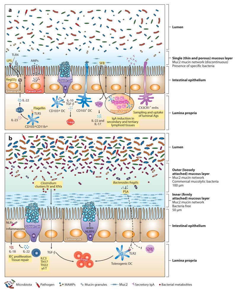
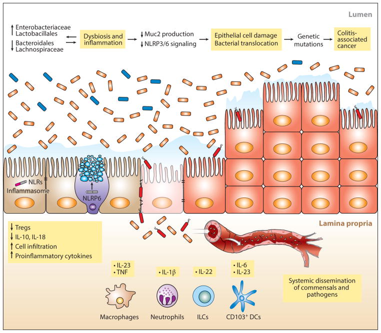
The diverse microbial populations constituting the intestinal microbiota promote immune development and differentiation, but because of their complex metabolic requirements and the consequent difficulty culturing them, they remained, until recently, largely uncharacterized and mysterious. In the last decade, deep nucleic acid sequencing platforms, new computational and bioinformatics tools, and full-genome characterization of several hundred commensal bacterial species facilitated studies of the microbiota and revealed that differences in microbiota composition can be associated with inflammatory, metabolic, and infectious diseases, that each human is colonized by a distinct bacterial flora, and that the microbiota can be manipulated to reduce and even cure some diseases. Different bacterial species induce distinct immune cell populations that can play pro- and anti-inflammatory roles, and thus the composition of the microbiota determines, in part, the level of resistance to infection and susceptibility to inflammatory diseases. This review summarizes recent work characterizing commensal microbes that contribute to the antimicrobial defense/inflammation axis.

Keywords: TLR signaling; antimicrobial molecules; commensals; dendritic cells; mucins.


Figures

Figure 1
Figure 1
Maintenance of intestinal homeostasis in the gastrointestinal tract. The intestinal epithelial surface is coated with a layer of mucus that has a pivotal role in intestinal barrier function. This mucous layer is organized by Muc2 mucin glycoproteins that polymerize into a gel-like structure, preventing luminal bacteria from coming into contact with epithelial cells. (b) In the large intestine, two mucous layers protect the colonic epithelium: a bacteria-free, dense, inner layer followed by an outer layer that harbors mucus-degrading bacteria. (a) In contrast, a single, loosely attached layer of mucus lines the small intestine. Mucins are produced by goblet cells and stored in secretory granules until appropriate stimulation, such as signaling through the Nod-like receptor pyrin domain 6 (NLRP6) inflammasome, prompts their release. Consistent with abundant mucus production in the colon, the number of goblet cells is much greater in the large intestine compared with the small bowel. The density and composition of luminal bacteria also differ between these two compartments. The small intestine harbors ~108 bacteria per gram of content. In mice, segmented filamentous bacteria (SFB) adhere to the intestinal surface and enhance the development of Th17 cells and the production of IgA by B cells. Microbial molecules stimulate the production of antimicrobial peptides (AMPs) from epithelial and Paneth cells through activation of innate immune receptors. For example, induction of regenerating islet-derived 3 γ(RegIIIγ), an antimicrobial protein that targets gram-positive bacteria and maintains host-bacterial segregation, is mediated through lipopolysaccharide (LPS) and flagellin stimulation of TLR4+ (Toll-like receptor 4) radioresistant cells and TLR5+CD103+CD11b+ dendritic cells (DCs), respectively. DCs and macrophages sample luminal antigens and stimulate cytokine production and T cell differentiation. Although CX3CR1+ macrophages are the major antigen-sampling mononuclear phagocytes in the small intestine, CD103+ DCs play a role as well. Nearly 1011–1012 bacteria reside in the large intestine. The microbiota are diverse, mainly comprising Lachnospiraceae, Bacteroides, and Clostridium groups IV and XIV. Tissue repair and tolerance to commensal and food antigens are key factors for maintaining homeostasis in the gut. IL-22 promotes epithelial cell proliferation and repair; however, excessive and aberrant signaling can lead to inflammation and colitis. Tolerance is accomplished by the induction of regulatory T cells (Tregs) through several mechanisms. In the small intestine, CD103+ DCs take up mucus and stimulate Tregs through IL-10 and TGF-β. Although the mechanism has not been elucidated, it is possible that DCs take up mucus directly by extending dendrites into the lumen or that goblet cells transfer mucus to DCs, as has been shown with antigen. In the colon, specific Treg-inducing bacteria have been identified. For instance, Clostridia spp. generate metabolites that upon receptor binding stimulate the production of TGF-βby epithelial cells. Polysaccharide A (PSA) from Bacteroides fragilis enhances Treg function (either through direct stimulation of TLR2+ Tregs or through a TLR2+ DC intermediate) while inhibiting proinflammatory Th17 responses. Abbreviations: ILC3, type 3 innate lymphoid cell; MAMPs, microbe-associated molecular patterns; mΦ, macrophage.
Figure 2
Figure 2
Inflammation, colitis, and cancer. Shifts in microbiota composition, in particular the expansion of γ-proteobacteria belonging to the Enterobacteriaceae family (orange), have been associated with colitis. These changes in bacterial composition are driven by factors such as infection, inflammation, antibiotic treatment, and deficiency of genes involved in the host-microbial response. Although specific bacteria that confer a colitogenic phenotype on normal hosts have been identified (188, 189), whether altered gut microbiota are the culprit behind colitis rather than a result of inflammation is still a matter of debate. Thinning of the mucous layer makes the intestine vulnerable to invasion by bacteria that may spread to other organs through the bloodstream. The continued presence of a colitogenic flora and production of proinflammatory cytokines plus alterations in the host genome perpetuate a state of inflammation that can eventually progress to cancer. Abbreviations: DC, dendritic cell; ILC, innate lymphoid cell; NLRP6, Nod-like receptor pyrin domain 6; Treg, T regulatory cell.

References

    1. Hejnol A, Martindale MQ. Acoel development indicates the independent evolution of the bilaterian mouth and anus. Nature. 2008;456:382–86. - PubMed
    1. Maxmen A. Evolution: a can of worms. Nature. 2011;470:161–62. - PubMed
    1. Honda K, Littman DR. The microbiome in infectious disease and inflammation. Annu Rev Immunol. 2012;30:759–95. - PMC - PubMed
    1. Belkaid Y, Hand TW. Role of the microbiota in immunity and inflammation. Cell. 2014;157:121–41. - PMC - PubMed
    1. Hum. Microbiome Proj. Consort. Structure, function and diversity of the healthy human microbiome. Nature. 2012;486:207–14. - PMC - PubMed

MeSH terms