. 2015 May;135(5):1425-1434.
doi: 10.1038/jid.2015.3. Epub 2015 Jan 12.

Kruppel-like factor KLF4 facilitates cutaneous wound healing by promoting fibrocyte generation from myeloid-derived suppressor cells

Affiliations

Kruppel-like factor KLF4 facilitates cutaneous wound healing by promoting fibrocyte generation from myeloid-derived suppressor cells

Lingling Ou et al. J Invest Dermatol. 2015 May.

Abstract

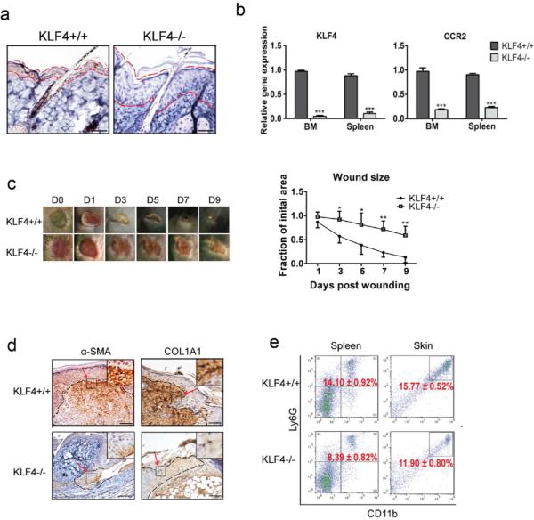
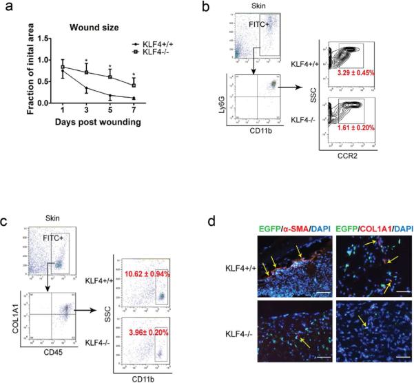
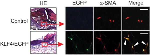
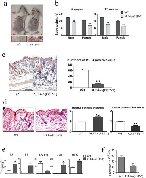
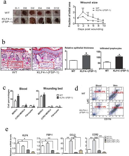
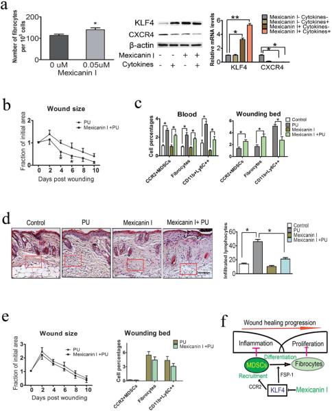
Pressure ulcers (PUs) are serious skin injuries whereby the wound healing process is frequently stalled in the inflammatory phase. Myeloid-derived suppressor cells (MDSCs) accumulate as a result of inflammation and promote cutaneous wound healing by mechanisms that are not fully understood. Recently, MDSCs have been shown to differentiate into fibrocytes, which serve as emerging effector cells that enhance cell proliferation in wound healing. We postulate that in wound healing MDSCs not only execute their immunosuppressive function to regulate inflammation but also stimulate cell proliferation once they differentiate into fibrocytes. In the current study, by using full-thickness and PU mouse models, we found that Kruppel-like factor 4 (KLF4) deficiency resulted in decreased accumulation of MDSCs and fibrocytes, and wound healing was significantly delayed. Conversely, KLF4 activation by the plant-derived product Mexicanin I increased the number of MDSCs and fibrocytes and accelerated the wound healing. Collectively, our study revealed a previously unreported function of MDSCs in cutaneous wound healing and identified Mexicanin I as a potential agent to accelerate PU wound healing.


Figures

Figure 1
Figure 1. KLF4 ablation delayed cutaneous wound healing accompanied by decreased accumulation of MDSCs
(a). KLF4 IHC staining in skin of RosaCreER/KLF4(flox) mice without (KLF4+/+) and with tamoxifen induction (KLF4−/−). The areas between two dotted red lines delineate skin suprabasal layers. (b). qRT-PCR to analyze expression of KLF4 and CCR2. (c). Wound healing phenotypes (left, n=10) and the wound size quantification (right). (d). IHC staining of α–SMA and COL1A1 in KLF4+/+ and KLF4−/− mice at Day 3 after wound induction. (e). Flow cytometry using anti-Ly6G and anti-CD11b antibodies with spleen cells and single skin cells. Representative images from one of five mice in each group are shown (a, c, and d). Scale bars: 50 μm. Mean ± SEM. *p< 0.05, **p< 0.01, ***p< 0.001.
Figure 2
Figure 2. Delayed wound healing in bone marrow KLF4 knockout mice and compromised accumulation of CCR2+MDSCs and fibrocytes
(a). Chimeric mice receiving bone marrow cells from Rosa26CreER/KLF4(flox)/β-actin-EGFP donor mice were used. Quantification of the wound size in each group of mice is shown (n=10). (b). Single cells from the skin wound were gated by EGFP. They were examined by CD11b and Ly6G antibodies, followed by further analysis using a CCR2 antibody. Representative contour plots in each group are shown. (c). Similar to (b) except COL1A1, CD45, and CD11b antibodies were used to analyze the fibrocytes. (d). Representative immunofluorescent staining of the wounds with α-SMA and COL1A1 antibodies. Yellow arrows indicate EGFP/α-SMA or EGFP/COL1A1 co-expressing cells (Scale bars: 50 μm).
Figure 3
Figure 3. KLF4-expressing bone marrow cells are integrated into the healing tissue and co-localized with α-SMA-expressing cells
Chimeric mice were generated by bone marrow transplantation using bone marrow cells from C57BL/6 mice (Control) or KLF4/EGFP mice into C57BL/6 recipient mice. 8mm diameter full thickness wound was placed and the wound beds were collected 4 days later followed by immunofluorescence staining with an anti-α-SMA antibody. Representative co-localization of EGFP cells with red α-SMA-expressing cells in the healing tissue (left) are indicated by white arrowheads (n=5). Scale bars: 25 μm.
Figure 4
Figure 4. Hair loss and decreased fibrocyte generation in FSP-1-Cre/KLF4(flox) mice
(a). Representatives of the wild type (WT) and FSP=1-Cre/KLF4(flox) (KLF4−/−(FSP-1)) mice. Black squares indicate an area in which a severe hair loss was seen in KLF4−/−(FSP-1) mice. (b). Body weights of male and female WT and KLF4−/−(FSP-1) mice. (c). Representative images of KLF4 staining of skin (left) and measurement of KLF4 positive cells (right). (d). Left, representative images of HE staining of skin. The areas between two dotted red lines represent skin suprabasal layers and the red arrow heads are pointing to hair follicles. Right, measurement of epithelial thickness and hair follicles. (e). Measurement of serum cytokines by ELISA (n=3). (f). Quantification of fibrocyte generation (n=5) Scale bars: 100 μm. *p<0.05, **p<0.01.
Figure 5
Figure 5. Compromised wound healing of PU in FSP-1-Cre/KLF4(flox) mice associated with decreased CCR2+MDSCs and fibrocytes
(a). Similar to Figure 1c, except the WT and KLF4−/−(FSP-1) mice were used in the PU model (n=10). (b). Left: Representative images of HE staining of skin wounds in PU model. Right: Quantification of the epithelial thickness and the numbers of infiltrated lymphocytes in arbitrary red squares with the same sizes as the left (n=5). (c). Flow cytometry analysis to examine MDSCs and fibrocytes in mouse blood and skin wounds. (d). Similar to c, CD11b and Ly6C antibodies were used to examine inflammatory monocytes. (e). qRT=PCR to analyze expression of KLF4, FSP-1, CCL2, and CCR2 in the skin wounds. Scale bars: 100 μm, *p<0.05, **p<0.01.
Figure 6
Figure 6. KLF4 activation by Mexicanin I accelerated PU wound healing accompanied by increased CCR2+MDSCs and fibrocytes
(a). Fibrocytes generation in the presence of 50 nM Mexicanin I (left), KLF4 expression detected by Western blotting (middle) and qRT=PCR (right). CXCR4 and β-actin were used as controls. (b). Measurement of wound healing kinetics after Mexicanin I treatment (n=10). (c): Flow cytometry analysis to examine different cell types. (d). Representatives of HE staining of the skin wounds (left) and numbers of infiltrated lymphocytes in the arbitrary squares with the same sizes from the left (right). (e). Left: measurement of wound healing kinetics in KLF4−/−(FSP-1) mice (n=10) and Right: flow cytometry analysis. (f). Proposed function of KLF4-mediated fibrocyte generation in wound healing. Scale bar: 100 μm, *p<0.05.

References

    1. Ai W, Zheng H, Yang X, et al. Tip60 functions as a potential corepressor of KLF4 in regulation of HDC promoter activity. Nucleic Acids Res. 2007;35:6137–49. - PMC - PubMed
    1. Alder JK, Georgantas RW, 3rd, Hildreth RL, et al. Kruppel-like factor 4 is essential for inflammatory monocyte differentiation in vivo. J Immunol. 2008;180:5645–52. - PMC - PubMed
    1. Ansel KM, Djuretic I, Tanasa B, et al. Regulation of Th2 differentiation and Il4 locus accessibility. Annu Rev Immunol. 2006;24:607–56. - PubMed
    1. Black JM, Edsberg LE, Baharestani MM, et al. Pressure ulcers: avoidable or unavoidable? Results of the National Pressure Ulcer Advisory Panel Consensus Conference. Ostomy Wound Manage. 2011;57:24–37. - PubMed
    1. Bujnicki T, Wilczek C, Schomburg C, et al. Inhibition of Myb-dependent gene expression by the sesquiterpene lactone mexicanin-I. Leukemia. 2012;26:615–22. - PubMed

Publication types

MeSH terms

LinkOut - more resources