Multicenter Study
. 2015 Mar;28(3):306-16.
doi: 10.1002/nbm.3252. Epub 2015 Jan 8.

Multi-center reproducibility of neurochemical profiles in the human brain at 7 T

Affiliations
Multicenter Study

Multi-center reproducibility of neurochemical profiles in the human brain at 7 T

B L van de Bank et al. NMR Biomed. 2015 Mar.

Abstract

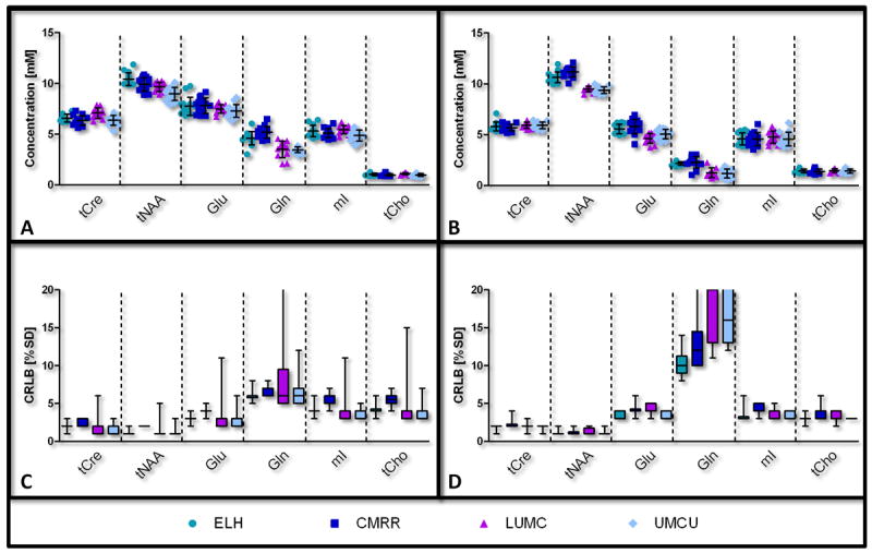
The purpose of this work was to harmonize data acquisition and post-processing of single voxel proton MRS ((1) H-MRS) at 7 T, and to determine metabolite concentrations and the accuracy and reproducibility of metabolite levels in the adult human brain. This study was performed in compliance with local institutional human ethics committees. The same seven subjects were each examined twice using four different 7 T MR systems from two different vendors using an identical semi-localization by adiabatic selective refocusing spectroscopy sequence. Neurochemical profiles were obtained from the posterior cingulate cortex (gray matter, GM) and the corona radiata (white matter, WM). Spectra were analyzed with LCModel, and sources of variation in concentrations ('subject', 'institute' and 'random') were identified with a variance component analysis. Concentrations of 10-11 metabolites, which were corrected for T1 , T2 , magnetization transfer effects and partial volume effects, were obtained with mean Cramér-Rao lower bounds below 20%. Data variances and mean concentrations in GM and WM were comparable for all institutions. The primary source of variance for glutamate, myo-inositol, scyllo-inositol, total creatine and total choline was between subjects. Variance sources for all other metabolites were associated with within-subject and system noise, except for total N-acetylaspartate, glutamine and glutathione, which were related to differences in signal-to-noise ratio and in shimming performance between vendors. After multi-center harmonization of acquisition and post-processing protocols, metabolite concentrations and the sizes and sources of their variations were established for neurochemical profiles in the healthy brain at 7 T, which can be used as guidance in future studies quantifying metabolite and neurotransmitter concentrations with (1) H-MRS at ultra-high magnetic field.

Keywords: 7 T; MRS; neurochemical profiling; reproducibility; semi-LASER; test-retest; ultra-high field strength.


Figures

Figure 1
Figure 1
In vivo 1H MR spectra of a single subject obtained using four different 7T systems for each scan & rescan from the two volumes of interest (VOI) using the semi-LASER sequence. All spectra are normalized to NAA (2.01ppm). Left (A&C): localization of the VOI at the Posterior Cingulate Cortex and the spectra that were obtained from an 8cc voxel (primarily gray matter). Right (B&D): localization of the VOI at the Corona Radiata in the left hemisphere and the resulting spectra from a 5.8cc voxel (primarily white matter). Note that high quality spectra could be obtained at all institutes.
Figure 2
Figure 2
The pulse sequence diagram of the semi-LASER used in the current study (adapted from (9), reprinted with permission). To suppress the water signal an 8-pulse VAPOR scheme was used. To suppress unwanted signals from outside the region of interest outer volume suppression (OVS) was used, consisting of a few pairs of adiabatic pulses applied in different dimensions. The voxel is selected by an asymmetric excitation pulse followed by two pairs of adiabatic full passage pulses (AFP) to refocus the signal. Based on a maximum achievable transmit B1 of 23μT for both locations on all systems the following parameters could be used: the duration of the VAPOR water suppression pulses was 35ms (BW: ~0.5ppm, around water resonance), duration of OVS (hyperbolic secant; BW: 9.1kHz; played out in subadiabatic regime with nominal flip angle of 90 degrees) pulses was 4.6ms, duration of the asymmetric slice selective excitation pulse was 4.2ms (maximum amplitude at 3.57ms, bandwidth 3.7kHz, adapted from (11)), and duration of the adiabatic slice selective refocusing pulses was 4.5ms (bandwidth 5.27kHz). An echo time of 30ms and a repetition time of 8 seconds was used, which ensured that all measurements remained within SAR guidelines for all sites and that T1 saturation was avoided.
Figure 3
Figure 3
Results of metabolite fitting for the semi-LASER sequence (TE=30ms, TR=8s, 64 averages) in the posterior cingulate cortex. The contributions of alanine (Ala), aspartate (Asp), ascorbate/vitamin C (Asc), glycerophosphocholine (GPC), phosphocholine (PC), creatine (Cr), phosphocreatine (PCr), γ-aminobutyric acid (GABA), glucose (Glc), glutamine (Gln), glutamate (Glu), glycine (Gly), glutathione (GSH), myo-inositol (mI), lactate (Lac), N-acetylaspartyl (NAA), N-acetylaspartylglutamate (NAAG), phosphoethanolamine (PE), scyllo-inositol (Scyllo), taurine (Tau), macromolecules (MM) and baseline to the in vivo spectrum are shown.
Figure 4
Figure 4
Fitting precision by LCModel of the macromolecular profiles and baselines per institute. The mean (black line) and standard deviation (gray area) of each fitted macromolecular profile and each fitted baseline is plotted per institute. (A) Differences in fitting of the macromolecular profiles are visible per site, but variations within sites are relatively small. (B) The baselines were fitted quite consistently by LCModel within both MR manufacturers.
Figure 5
Figure 5
LCModel quantification results for selected metabolites per institute. The concentrations of the metabolites are shown as mean ± SD, as well as individual data points (A&B) and their corresponding Cramér-Rao Lower Bounds (CRLB) in box plots (C&D). Left: posterior cingulate cortex (A&C); right: corona radiata (B&D). Tissue levels of Cre+PCr (tCre), NAA+NAAG (tNAA), Glu, Gln, mI and tCho are shown. Note the minor deviations in total variance between the four contributing institutes. A list of abbreviations can be found in Figure 3.
Figure 6
Figure 6
Repeatability of metabolites (A&B) presented in Figure 5. The concentrations obtained from the first scan (horizontal axis) are plotted against the concentrations from the second scan (vertical axis), with left – posterior cingulate cortex and right – corona radiata. Note that the close proximity of these metabolites to the identity line (x=y) reveals high test-retest reproducibility, also shown by the low CoV. The bottom row (C&D) shows a zoomed-in view of the tCho concentrations. The coefficient of variation (CoV) for this metabolite was 4.0% for gray matter (ICCsubject = 57%) and 3.4% for white matter (ICCsubject = 85%). It is clear that most variation results from differences between volunteers (markers, clustered), rather than from differences between institutes (colors, more randomly distributed).

References

    1. Oz G, Alger JR, Barker PB, Bartha R, Bizzi A, Boesch C, Bolan PJ, Brindle KM, Cudalbu C, Dincer A, Dydak U, Emir UE, Frahm J, Gonzalez RG, Gruber S, Gruetter R, Gupta RK, Heerschap A, Henning A, Hetherington HP, Howe FA, Huppi PS, Hurd RE, Kantarci K, Klomp DW, Kreis R, Kruiskamp MJ, Leach MO, Lin AP, Luijten PR, Marjanska M, Maudsley AA, Meyerhoff DJ, Mountford CE, Nelson SJ, Pamir MN, Pan JW, Peet AC, Poptani H, Posse S, Pouwels PJ, Ratai EM, Ross BD, Scheenen TW, Schuster C, Smith IC, Soher BJ, Tkac I, Vigneron DB, Kauppinen RA. Clinical proton MR spectroscopy in central nervous system disorders. Radiology. 2014;270(3):658–679. - PMC - PubMed
    1. Duarte JM, Lei H, Mlynarik V, Gruetter R. The neurochemical profile quantified by in vivo 1H NMR spectroscopy. Neuroimage. 2012;61(2):342–362. - PubMed
    1. Ugurbil K, Adriany G, Andersen P, Chen W, Garwood M, Gruetter R, Henry PG, Kim SG, Lieu H, Tkac I, Vaughan T, Van De Moortele PF, Yacoub E, Zhu XH. Ultrahigh field magnetic resonance imaging and spectroscopy. Magnetic Resonance Imaging. 2003;21(10):1263–1281. - PubMed
    1. Tkac I, Oz G, Adriany G, Ugurbil K, Gruetter R. In vivo 1H NMR spectroscopy of the human brain at high magnetic fields: metabolite quantification at 4T vs. 7T. Magn Reson Med. 2009;62(4):868–879. - PMC - PubMed
    1. Emir UE, Auerbach EJ, Van De Moortele PF, Marjanska M, Ugurbil K, Terpstra M, Tkac I, Oz G. Regional neurochemical profiles in the human brain measured by (1)H MRS at 7 T using local B(1) shimming. NMR Biomed. 2012;25(1):152–160. - PMC - PubMed

Publication types