. 2015 Jun;145(2):242-57.
doi: 10.1111/imm.12440.

Immune response in the adipose tissue of lean mice infected with the protozoan parasite Neospora caninum

Affiliations

Immune response in the adipose tissue of lean mice infected with the protozoan parasite Neospora caninum

Luzia Teixeira et al. Immunology. 2015 Jun.

Abstract

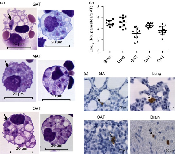
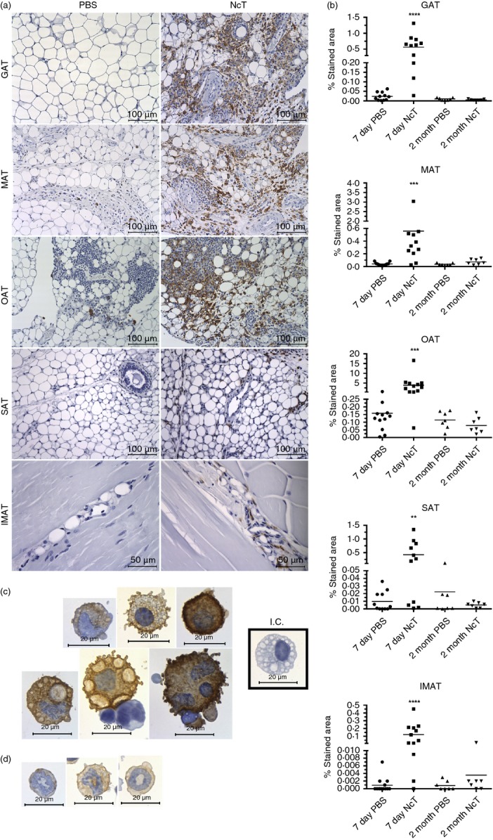
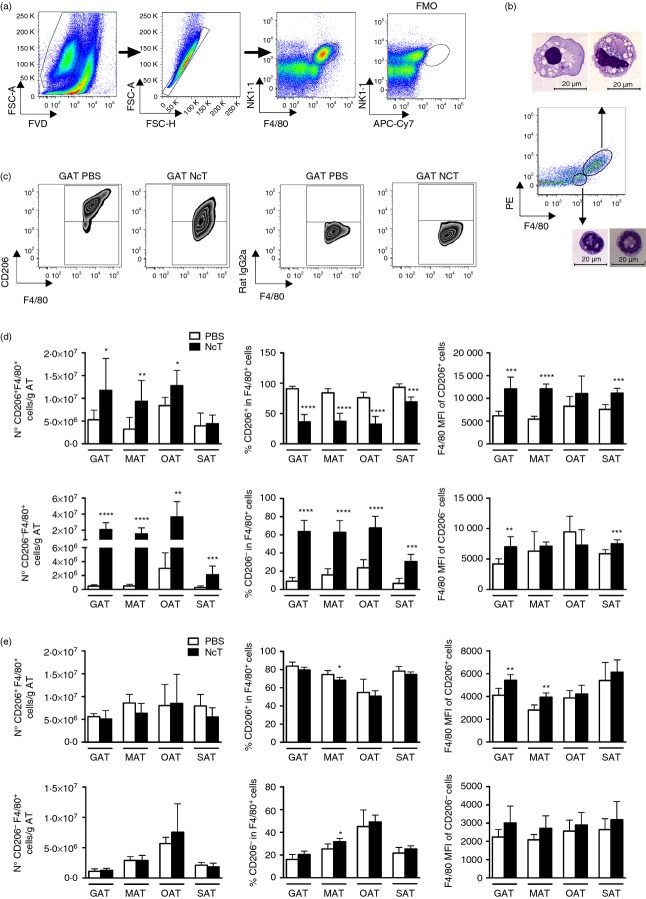
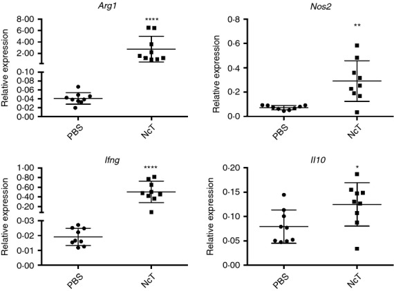
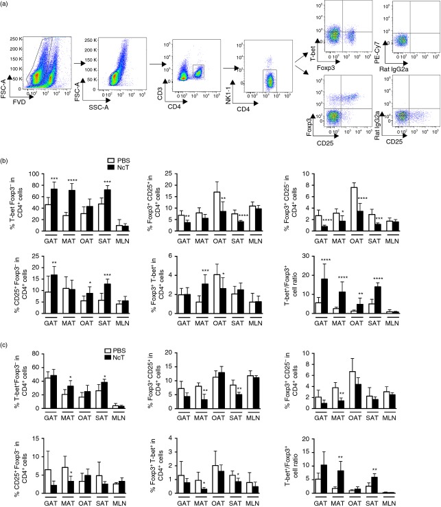
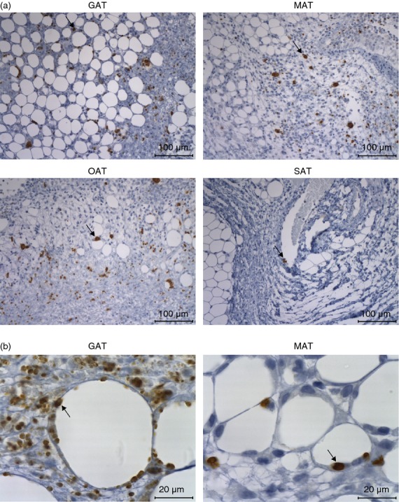
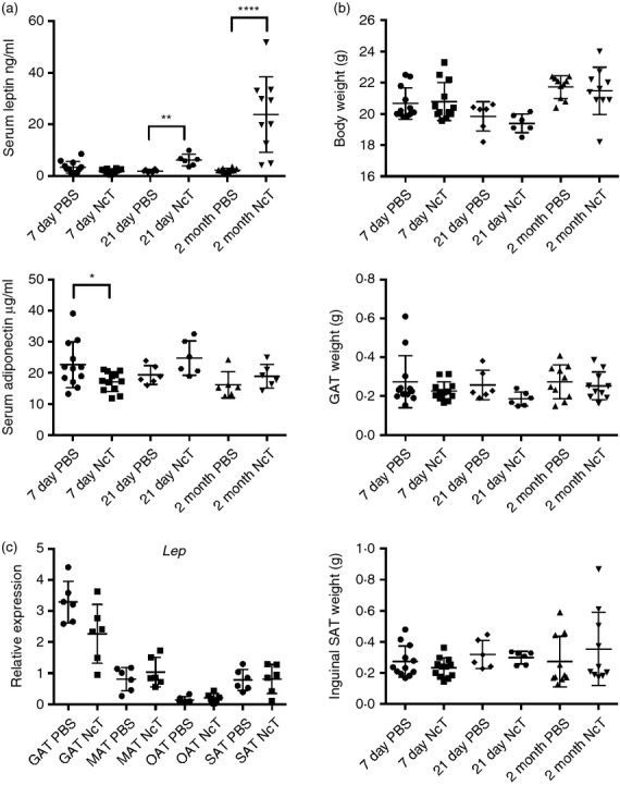
The adipose tissue can make important contributions to immune function. Nevertheless, only a limited number of reports have investigated in lean hosts the immune response elicited in this tissue upon infection. Previous studies suggested that the intracellular protozoan Neospora caninum might affect adipose tissue physiology. Therefore, we investigated in mice challenged with this protozoan if immune cell populations within adipose tissue of different anatomical locations could be differently affected. Early in infection, parasites were detected in the adipose tissue and by 7 days of infection increased numbers of macrophages, regulatory T (Treg) cells and T-bet(+) cells were observed in gonadal, mesenteric, omental and subcutaneous adipose tissue. Increased expression of interferon-γ was also detected in gonadal adipose tissue of infected mice. Two months after infection, parasite DNA was no longer detected in these tissues, but T helper type 1 (Th1) cell numbers remained above control levels in the infected mice. Moreover, the Th1/Treg cell ratio was higher than that of controls in the mesenteric and subcutaneous adipose tissue. Interestingly, chronically infected mice presented a marked increase of serum leptin, a molecule that plays a role in energy balance regulation as well as in promoting Th1-type immune responses. Altogether, we show that an apicomplexa parasitic infection influences immune cellular composition of adipose tissue throughout the body as well as adipokine production, still noticed at a chronic phase of infection when parasites were already cleared from that particular tissue. This strengthens the emerging view that infections can have long-term consequences for the physiology of adipose tissue.

Keywords: Neospora caninum; adipose tissue; leptin; macrophages; regulatory T cells.


Figures

Figure 1
Figure 1
Parasitic burden in the adipose tissue of infected mice. (a) Hemacolor staining of stromal vascular fraction cells isolated from gonadal adipose tissue (GAT), mesenteric adipose tissue (MAT) and omental adipose tissue (OAT) of C57BL/6 wild-type (WT) mice 6 hr after intraperitoneal administration of 1 × 107 Neospora caninum tachyzoites (NcT). Parasites are observed inside cells with macrophage-like morphology (indicated by arrows). (b) Parasitic load expressed as Log10 of the number of parasites per gram of tissue, determined by PCR analysis, of the indicated organs or adipose tissue of WT mice 7 days after NcT inoculation. Each symbol represents an individual mouse. Horizontal lines represent the mean values of the respective group (± SD). These are pooled results from three independent experiments. (c) Detection of N. caninum in the indicated tissues/organs of WT mice 7 days after NcT inoculation by immunohistochemistry analysis. Thin sections of the indicated organs/tissues were specifically stained (brown coloration, indicated by arrows) with a polyclonal serum goat anti-N. caninum and counterstained with haematoxylin.
Figure 2
Figure 2
Increased F4/80-stained area in the adipose tissue of Neospora caninum-infected mice. (a) Representative immunohistochemistry analysis of F4/80 in gonadal adipose tissue (GAT), mesenteric adipose tissue (MAT), omental adipose tissue (OAT), subcutaneous adipose tissue (SAT) and intramuscular adipose tissue (IMAT) from wild-type C57BL/6 mice 7 days after intraperitoneal administration of 1 × 10N. caninum tachyzoites (NcT) or PBS. Adipose tissue was specifically stained (brown coloration) with a monoclonal anti-mouse F4/80 antibody and counterstained with haematoxylin. (b) Frequency of stained area of analysed adipose tissue depots at 7 days and 2 months after challenge. Each symbol represents an individual mouse. Horizontal lines represent the mean values of the respective group. These are pooled results from three (7 days) or two (2 months) independent experiments. Statistically significant differences between different experimental groups are indicated (Mann–Whitney U-test, ** 0·01; *** 0·001; **** 0·0001). Immunohistochemistry analysis of F4/80 in stromal vascular fraction cells isolated from the GAT of infected C57BL/6 mice, killed 7 days after the parasitic challenge. Cells were specifically stained (brown coloration) with a monoclonal rat IgG2a anti-mouse F4/80 antibody and counterstained with haematoxylin. Cells with morphology compatible with macrophages (c) and polymorphonuclear cells (d) are observed. I.C. (Cells stained with Isotype Control, Rat IgG2a). Bar = 100 µm in all micrographs but IMAT where bar = 50 µm.
Figure 3
Figure 3
Increased macrophage numbers in the adipose tissue of Neospora caninum-infected mice. (a) Flow cytometry gating strategy used to define macrophages in the stromal vascular fraction (SVF) of the different depots of adipose tissue analysed. Dead cells were excluded with Fixable Viability Dye (FVD) and Singlets were then selected from FSC-A versus FSC-H dot plot. Macrophages were defined as F4/80high autofluorescencehigh. The fluorescence minus one (FMO) control is also shown. Dot plots are representative examples with SVF cells isolated from gonadal adipose tissue (GAT). (b) Hemacolor staining of sorted F4/80high autofluorescencehigh and F4/80low SVF cells, as indicated, from GAT of mice 7 days after intraperitoneal administration of 1 × 10N. caninum tachyzoites. Bar = 20 µm in all micrographs (c) Representative zebra plots of CD206+ F4/80+ and CD206 F4/80+ SVF cells gated in F4/80high autofluorescencehigh cells from of GAT of mice 7 days after parasitic challenge (NcT) or PBS (PBS). Respective isotype controls are shown. Numbers of CD206+ F4/80+ or CD206 F4/80+ cells per gram of adipose tissue or frequencies of the same cell populations in F4/80+ cells in the gonadal adipose tissue (GAT), mesenteric adipose tissue (MAT), omental adipose tissue (OAT) and subcutaneous adipose tissue (SAT) at (d) 7 days and (e) 2 months after intraperitoneal challenge with 1 × 107 N. caninum tachyzoites (NcT) or PBS, as indicated. Mean fluorescence intensity (MFI) for F4/80 is also presented in the respective population of macrophages. Bars represent mean plus one SD of six to nine mice per group pooled from 3 (7 days) or 2 (2 months) independent experiments. (Mann–Whitney U-test, *< 0·05; ** 0·01; *** 0·001; **** 0·0001).
Figure 4
Figure 4
Increased expression of M1 and M2 macrophage markers in the adipose tissue of infected mice. Relative levels of arginase (Arg1), nitric oxide synthase 2, inducible (Nos2), interferon-γ (Ifng) and interleukin 10 (Il10) mRNA, normalized to Non-POU-domain containing octamer binding protein (Nono) mRNA, detected by real-time PCR in the stromal vascular fraction (SFV) of gonadal adipose tissue (GAT) of mice 7 days after intraperitoneal administration of 1 × 10Neospora caninum tachyzoites (NcT) or PBS (PBS). Each symbol represents an individual mouse. Horizontal lines represent the mean values of the respective group (± SD). These results are pooled from three independent experiments. (Mann–Whitney U-test, *< 0·05; ** 0·01; **** 0·0001).
Figure 5
Figure 5
Increased T helper type 1 (Th1)/regulatory T (Treg) cell ratio in the adipose tissue of Neospora caninum-infected mice. (a) Flow cytometry analysis of Th1 cells (defined as T-bet+ Foxp3 CD4+ CD3+ NK) and Treg cells in stromal vascular fraction cells isolated from omental adipose tissue. Dot plots are representative examples of gate strategy used to define Th1 and Treg cells in the different depots of adipose tissue analysed. Dead cells were excluded with Fixable Viability Dye (FVD). Percentages of T-bet+ Foxp3 cells, Foxp3+ CD25+ cells, Foxp3+ CD25 cells, CD25+ Foxp3 cells and Foxp3+ T-bet+ on total CD4+ CD3+ NK cells in the gonadal adipose tissue (GAT), mesenteric adipose tissue (MAT), omental adipose tissue (OAT), subcutaneous adipose tissue (SAT) and mesenteric lymph nodes (MLN) from C57BL/6 mice killed 7 days (b) and 2 months (c) after intraperitoneal challenge with 1 × 107 N. caninum tachyzoites (NcT) or PBS, as indicated. The ratio of Th1 cells (T-bet+ Foxp3 CD4+) and total Treg cells (Foxp3+ T-bet CD4+) is also depicted for the different adipose tissue depots analysed. These are pooled results from three (7 days) or two (2 months) independent experiments. Bars represent means plus one SD of six to nine animals per group. (Mann–Whitney U-test, *< 0·05; ** 0·01; *** 0·001; **** 0·0001).
Figure 6
Figure 6
Detection of Neospora caninum in the adipose tissue of infected interleukin-12 (IL-12)/IL-23 p40−/− mice. (a) Representative images showing parasitic forms in the gonadal adipose tissue (GAT), mesenteric adipose tissue (MAT), omental adipose tissue (OAT) and subcutaneous adipose tissue (SAT) of p40−/− C57BL/6 mice 7 days after intraperitoneal administration of 5 × 105 N. caninum tachyzoites, detected by immunohistochemistry. Thin sections of the indicated organs/tissues were specific stained (brown coloration, indicated by arrows) with a polyclonal serum goat anti-N. caninum and counterstained with haematoxylin. This is one representative result of two independent experiments with three mice per experiment. Bar = 100 µm (b) Higher magnification of GAT and MAT showing parasitic forms closely associated with adipocytes (arrow). Bar = 20 µm.
Figure 7
Figure 7
Leptin levels increase in the serum of Neospora caninum-infected mice. (a) Serum levels of leptin and adiponectin and (b) total body, gonadal adipose tissue (GAT) and inguinal subcutaneous adipose tissue (SAT) weight, as indicated, of C57BL/6 mice 7 days, 21 days and 2 months after intraperitoneal administration of 1 × 10N. caninum tachyzoites (NcT) or PBS. (c) Relative levels of leptin (Lep) mRNA, normalized to Non-POU-domain containing octamer binding protein (Nono) mRNA, detected by real-time PCR in GAT, mesenteric adipose tissue (MAT), omental adipose tissue (OAT) and SAT of mice 2 months after intraperitoneal administration of 1 × 107 NcT or PBS. Each symbol represents an individual mouse. Horizontal lines represent the mean values of the respective group (± SD). These are pooled results from two to three independent experiments. (Mann–Whitney U-test, *< 0·05; ** 0·01; **** 0·0001).

References

    1. Schaffler A, Scholmerich J. Innate immunity and adipose tissue biology. Trends Immunol. 2010;31:228–35. - PubMed
    1. Mathis D. Immunological goings-on in visceral adipose tissue. Cell Metab. 2013;17:851–9. - PMC - PubMed
    1. Nagajyothi F, Desruisseaux MS, Machado FS, et al. Response of adipose tissue to early infection with Trypanosoma cruzi (Brazil strain) J Infect Dis. 2012;205:830–40. - PMC - PubMed
    1. Gray KS, Collins CM, Speck SH. Characterization of omental immune aggregates during establishment of a latent gammaherpesvirus infection. PLoS One. 2012;7:e43196. - PMC - PubMed
    1. Na HN, Nam JH. Adenovirus 36 as an obesity agent maintains the obesity state by increasing MCP-1 and inducing inflammation. J Infect Dis. 2012;205:914–22. - PubMed

Publication types

MeSH terms

LinkOut - more resources