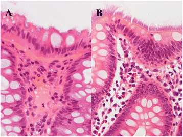
Clinicopathologic study of intestinal spirochetosis in Japan with special reference to human immunodeficiency virus infection status and species types: analysis of 5265 consecutive colorectal biopsies
- PMID: 25582884
- PMCID: PMC4300994
- DOI: 10.1186/s12879-014-0736-4
Clinicopathologic study of intestinal spirochetosis in Japan with special reference to human immunodeficiency virus infection status and species types: analysis of 5265 consecutive colorectal biopsies
Abstract
Background: Previous studies reported that the incidence of intestinal spirochetosis was high in homosexual men, especially those with Human Immunodeficiency Virus infection. The aim of the present study was to clarify the clinicopathological features of intestinal spirochetosis in Japan with special reference to Human Immunodeficiency Virus infection status and species types.
Methods: A pathology database search for intestinal spirochetosis was performed at Tokyo Metropolitan Cancer and Infectious Disease Center Komagome Hospital between January 2008 and October 2011, and included 5265 consecutive colorectal biopsies from 4254 patients. After patient identification, a retrospective review of endoscopic records and clinical information was performed. All pathology slides were reviewed by two pathologists. The length of the spirochetes was measured using a digital microscope. Causative species were identified by polymerase chain reaction.
Results: Intestinal spirochetosis was diagnosed in 3 out of 55 Human Immunodeficiency Virus-positive patients (5.5%). The mean length of intestinal spirochetes was 8.5 μm (range 7-11). Brachyspira pilosicoli was detected by polymerase chain reaction in all 3 patients. Intestinal spirochetosis was also diagnosed in 73 out of 4199 Human Immunodeficiency Virus-negative patients (1.7%). The mean length of intestinal spirochetes was 3.5 μm (range 2-8). The species of intestinal spirochetosis was identified by polymerase chain reaction in 31 Human Immunodeficiency Virus-negative patients. Brachyspira aalborgi was detected in 24 cases (78%) and Brachyspira pilosicoli in 6 cases (19%). Both Brachyspira aalborgi and Brachyspira pilosicoli were detected in only one Human Immunodeficiency Virus-negative patient (3%). The mean length of Brachyspira aalborgi was 3.8 μm, while that of Brachyspira pilosicoli was 5.5 μm. The length of Brachyspira pilosicoli was significantly longer than that of Brachyspira aalborgi (p < 0.01). The lengths of intestinal spirochetes were significantly longer in Human Immunodeficiency Virus-positive patients than in Human Immunodeficiency Virus-negative patients (p < 0.05).
Conclusions: The incidence of intestinal spirochetosis was slightly higher in Human Immunodeficiency Virus-positive patients than in Human Immunodeficiency Virus-negative patients. However, no relationship was found between the Human Immunodeficiency Virus status and intestinal spirochetosis in Japan. Brachyspira pilosicoli infection may be more common in Human Immunodeficiency Virus-positive patients with intestinal spirochetosis than in Human Immunodeficiency Virus-negative patients with intestinal spirochetosis.
Figures

References
-
- Smith JL. Colonic spirochetosis in animals and humans. J Food Prot. 2005;68(7):1525–1534. - PubMed
-
- Takeuchi A, Jervis HR, Nakazawa H, Robinson DM. Spiral-shaped organisms on the surface colonic epithelium of the monkey and man. Am J Clin Nutr. 1974;27(11):1287–1296. - PubMed
-
- Stephens CP, Hampson DJ. Intestinal spirochete infections of chickens: a review of disease associations, epidemiology and control. Anim Health Res Rev. 2001;2(1):83–91. - PubMed
-
- Esteve M, Salas A, Fernandez-Banares F, Lloreta J, Marine M, Gonzalez CI, Forne M, Casalots J, Santaolalla R, Espinos JC, et al. Intestinal spirochetosis and chronic watery diarrhea: clinical and histological response to treatment and long-term follow up. J Gastroenterol Hepatol. 2006;21(8):1326–1333. doi: 10.1111/j.1440-1746.2006.04150.x. - DOI - PubMed
MeSH terms
Substances
LinkOut - more resources
Full Text Sources
Other Literature Sources
Medical

