. 2015 May;20(3):411-20.
doi: 10.1007/s12192-014-0565-9. Epub 2015 Jan 13.

MicroRNA-1 aggravates cardiac oxidative stress by post-transcriptional modification of the antioxidant network

Affiliations

MicroRNA-1 aggravates cardiac oxidative stress by post-transcriptional modification of the antioxidant network

Lu Wang et al. Cell Stress Chaperones. 2015 May.

Abstract

Oxidative stress plays an important role in cardiovascular diseases. Studies have shown that miR-1 plays an important role in the regulation of cardiomyocyte apoptosis, which can be the result of oxidative stress. This study was designed to determine whether increased miR-1 levels lead to alterations in the expression of proteins related to oxidative stress, which could contribute to heart dysfunction. We compared cardiac function in wild-type (WT) and miR-1 transgene (miR-1/Tg) C57BL/6 mice (n ≥ 10/group). Echocardiography showed that stroke volume (SV), ejection fraction (EF), and fractional shortening (FS) were significantly decreased in miR-1/Tg mice. Concomitantly, the level of reactive oxygen species (ROS) was elevated in the cardiomyocytes from the miR-1/Tg mice, and activities of lactate dehydrogenase (LDH) and creatinine kinase (CK) in plasma were also increased in the miR-1/Tg mice. All of these changes could be reversed by LNA-anti-miR-1. In the cardiomyocytes of neonatal Wistar rats, overexpression of miR-1 exhibits higher ROS levels and lower resistance to H2O2-induced oxidative stress. We demonstrated that SOD1, Gclc, and G6PD are novel targets of miR-1 for post-transcriptional repression. MicroRNA-1 post-transcriptionally represses the expression of SOD1, Gclc, and G6PD, which is likely to contribute to the increased ROS level and the susceptibility to oxidative stress of the hearts of miR-1 transgenic mice.


Figures
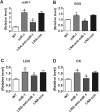
Fig. 1
Fig. 1
MiR-1 LNA reversed the degree of cardiac damage in miR-1 mice. MiR-1 LNA reduced ROS levels and the degree of cardiac damage in miR-1 mice. a MiR-1 levels were significantly increased in the hearts of miR-1 transgenic mice compared with those of normal controls, and miR-1 LNA decreased miR-1 levels in the hearts of miR-1 transgenic mice, while LNA-con had no effect on miR-1 levels. b ELISA results showed that the ROS level was increased in the hearts of miR-1 transgenic mice compared with the normal control, and miR-1 LNA decreased ROS levels in the hearts of miR-1 transgenic mice, while LNA-con did not decrease ROS levels. c LDH activity was increased in the hearts of miR-1 transgenic mice compared with those of the normal controls, and miR-1 LNA decreased LDH activity in the hearts of miR-1 transgenic mice, while LNA-con had no effect on LDH activity. d CK activity was increased in the hearts of miR-1 transgenic mice compared with those of the normal controls, and miR-1 LNA decreased CK activity in the hearts of miR-1 transgenic mice, while LNA-con did not decrease CK activity. Mean ± SEM. n = 6 mice in each group. *P < 0.05 versus WT; # P < 0.05 versus WT miR-1; unpaired Student’s t test
Fig. 2
Fig. 2
MiR-1-overexpressed cardiomyocytes exhibit higher ROS levels and lower resistance to oxidative stress. Cardiomyocytes were treated with miR-con, miR-1, miR-1, and AMO-1 or miR-1 and AMO-con for 24 h, and ROS levels were assessed by flow cytometry. a Representative results of cardiomyocytes that were treated with miR-con, miR-1, miR-1, and AMO-1 or miR-1 and AMO-con for 24 h and the ROS levels that were detected by flow cytometry analysis. b The fluorescence intensity was significantly increased in cardiomyocytes with miR-1 overexpression. AMO-1 decreased the fluorescence intensity significantly in cardiomyocytes with miR-1 overexpression, while AMO-con did not display the effect. Cardiomyocytes were treated with miR-con, miR-1, miR-1, and AMO-1 or miR-1 and AMO-con for 24 h and then treated with H2O2 (100 μM) for 12 h. Representative images of cardiomyocytes before c and after d treatment with H2O2. The cell pictures are magnified by ×100. e, f Quantification of cell viability. *P < 0.05
Fig. 3
Fig. 3
Verification of SOD1 (a), Gclc (b), and G6PD (c) as cognate targets of miR-1 for post-transcriptional repression. Verification of interactions between rat miR-1 and the wild or mutant 3′-UTRs of rat SOD1, Gclc, and G6PD in HEK293 cells is determined by luciferase reporter activity. Cells were transfected with the SOD1, Gclc, or G6PD construct as indicated. MiR-1 repressed luciferase reporter gene activity with the WT 3′-UTR of SOD1, Gclc, or G6PD. However, miR-1 failed to affect luciferase activity with the MT 3′-UTR of SOD1, Gclc, or G6PD. AMO-1 reversed miR-1 induced repression of luciferase reporter gene activity, but AMO-con did not display the effect. Mean ± SEM; n = 8 batches of cells in each group; *p < 0.05 versus miR-con; # p < 0.05 versus WT miR-1; unpaired Student’s t test
Fig. 4
Fig. 4
Effect of miR-1 on the mRNA and protein expression of antioxidant proteins in miR-1 transgenic mouse hearts under oxidative stress in vivo (with I/R perfusion). a The protein expression of Gclc, SOD1, and G6PD in miR-1 transgenic mouse hearts. The protein expression of Gclc, SOD1, and G6PD were not affected under normal conditions. I/R perfusion increased these protein expressions, but miR-1 attenuated the increase. Western blot analyses were repeated three times, and this figure is representative of only one analysis. b The mRNA expression of Gclc, SOD1, and G6PD in wild-type control mice and in miR-1 transgenic mice after suffering I/R perfusion. The mRNA expressions of Gclc, SOD1, and G6PD did not change in miR-1 transgenic mouse hearts after the transgenic mice suffered I/R perfusion. n = 6 mice in each group; *P < 0.05 versus WT; # P < 0.05 versus I/R; unpaired Student’s t test
Fig. 5
Fig. 5
Effect of miR-1 on the mRNA and protein expression of antioxidant proteins in miR-1-overexpressed rat ventricular cardiomyocytes under oxidative stress in vitro (stimulated with H2O2). a The expression of Gclc, SOD1, and G6PD at the protein level in neonatal rat ventricular cardiomyocytes. Cardiomyocytes were treated with miR-con, miR-1, or MT miR-1 for 24 h and then treated with H2O2 (100 μM) for 14 h. The protein expression of Gclc, SOD1, and G6PD was then assessed. MiR-1 did not affect the protein expression of Gclc, SOD1, and G6PD, and these protein expressions did not change in miR-1-treated cardiomyocytes after treatment with H2O2 (100 μM) for 14 h. Western blot analyses were repeated three times, and this figure is representative of only one analysis. b The expression of Gclc, SOD1, and G6PD at the mRNA level in neonatal rat ventricular cardiomyocytes treated with miR-con, miR-1, or MT miR-1 for 24 h, followed by treatment with H2O2 (100 μM) for 14 h. The mRNA expressions of Gclc, SOD1, and G6PD did not change in miR-1-treated cardiomyocytes, and these mRNA expressions did not change in miR-1-treated cardiomyocytes after stimulation with H2O2.*P < 0.05 versus miR-con; # p < 0.05 versus H2O2 + miR-con; unpaired Student’s t test

References

    1. Ai J, Zhang R, Li Y, Pu J, Lu Y, Jiao J, Li K, Yu B, Li Z, Wang R, et al. Circulating microRNA-1 as a potential novel biomarker for acute myocardial infarction. Biochem Biophys Res Commun. 2010;391(1):73–77. doi: 10.1016/j.bbrc.2009.11.005. - DOI - PubMed
    1. Anderson ME, Mohler PJ. MicroRNA may have macro effect on sudden death. Nat Med. 2007;13(4):410–411. doi: 10.1038/nm0407-410. - DOI - PubMed
    1. Chen Z, Oberley TD, Ho Y, Chua CC, Siu B, Hamdy RC, Epstein CJ, Chua BH. Overexpression of CuZnSOD in coronary vascular cells attenuates myocardial ischemia/reperfusion injury. Free Radic Biol Med. 2000;29(7):589–596. doi: 10.1016/S0891-5849(00)00363-4. - DOI - PubMed
    1. Chen T, Ding G, Jin Z, Wagner MB, Yuan Z. Insulin ameliorates miR-1-induced injury in H9c2 cells under oxidative stress via Akt activation. Mol Cell Biochem. 2012;369(1–2):167–174. doi: 10.1007/s11010-012-1379-7. - DOI - PubMed
    1. Cheng X, Siow RC, Mann GE. Impaired redox signaling and antioxidant gene expression in endothelial cells in diabetes: a role for mitochondria and the nuclear factor-E2-related factor 2-Kelch-like ECH-associated protein 1 defense pathway. Antioxid Redox Signal. 2011;14(3):469–487. doi: 10.1089/ars.2010.3283. - DOI - PubMed

Publication types

MeSH terms