. 2015 Jan 13:6:5981.
doi: 10.1038/ncomms6981.

Coherent coupling of molecular resonators with a microcavity mode

Affiliations

Coherent coupling of molecular resonators with a microcavity mode

A Shalabney et al. Nat Commun. .

Abstract

The optical hybridization of the electronic states in strongly coupled molecule-cavity systems have revealed unique properties, such as lasing, room temperature polariton condensation and the modification of excited electronic landscapes involved in molecular isomerization. Here we show that molecular vibrational modes of the electronic ground state can also be coherently coupled with a microcavity mode at room temperature, given the low vibrational thermal occupation factors associated with molecular vibrations, and the collective coupling of a large ensemble of molecules immersed within the cavity-mode volume. This enables the enhancement of the collective Rabi-exchange rate with respect to the single-oscillator coupling strength. The possibility of inducing large shifts in the vibrational frequency of selected molecular bonds should have immediate consequences for chemistry.


Figures

Figure 1
Figure 1. Polymer vibrational spectrum.
(a) Transmission spectrum of polyvinyl acetate (PVAc) thin layer deposited on a Ge substrate. The thickness of the film is about 2 μm and the measurement was performed at normal incidence. The measured transmission is normalized to free-space transmission. The black line fits the data modelling the polymer dispersion by ideal damped harmonic oscillators (see Supplementary Note 2). The inset shows the absorption band of PVAc due to the (C=O)-bond-stretching band around 1,740 cm−1 with the same fit (black line). (b) Chemical structure of a single PVAc monomer unit. (c) Three-dimensional structure of one PVAc monomer showing the (C=O) bond.
Figure 2
Figure 2. Microcavity and experimental set-up.
(a) Schematic illustration of the microcavity used to strongly couple the (C=O) vibrational band to IR radiation. A thin (~2 μm) layer of polyvinyl acetate (PVAc) is sandwiched between two symmetrical thin (10 nm ) Au mirrors deposited on a Ge substrate (see Supplementary Note 1). The (C=O) bonds are depicted as mechanical oscillators inside the cavity with arbitrary orientations. The red thick curve describes the electric field intensity spatial distribution for the first cavity mode tuned in resonance with the (C=O) vibrational transition. (b) Vibrational energy diagram in the anharmonic potential of the fundamental electronic state. The inset on the right-top shows the coupling scheme between the fundamental vibrational mode and the first optical mode ℏωc of the cavity and therefore forming two polariton branches (ℏωH and ℏωL ) separated by the Rabi energy. (c) The experimental set-up is based on a single-beam Fourier transform IR system that records on a DTGS (deuterated triglycine sulfate) detector an interferogram generated from a movable mirror (MM). BS indicates the beam-splitter. The interferogram is Fourier transformed to provide the actual vibrational spectrum. (d) Photographic image of a free-standing PVAc layer, clearly showing the continuous character of the film. (e) Photographic image of the cavity used in the experiments.
Figure 3
Figure 3. Cavity angular dispersion and strong coupling.
(a) Cascade plot of measured transmission spectra through the Au-polyvinyl acetate (PVAc) cavity as a function of the IR-beam incidence angle. The spectra are vertically shifted every 5 degrees and the angular range covers −60; +60 degrees relatively to the cavity normal. At normal incidence (θ=0°), the avoided crossing is clearly revealed as the signature of the strong coupling regime between the cavity mode and the (C=O) stretching mode (which position in an uncoupled situation is indicated by the vertical line). (b) Colour plot of the cavity (Au-PVAc) dispersion calculated with parameters retrieved from the best transmission data fit at normal incidence (see Supplementary Note 2). White diamonds and purple circles correspond, respectively, to the measured positions of the upper (UP) and lower (LP) polaritons extracted from the data displayed in a. Dashed curve and dashed horizontal line show, respectively, the dispersion of the empty cavity and (C=O) vibrational mode (see Supplementary Note 2). The dispersion of the empty cavity was calculated by deactivating vibrational contributions and considering the background refractive index of the polymer. The crossing point between the dashed curves at normal incidence corresponds to the careful tuning between the first mode of the cavity with the (C=O)-bond-stretching mode. The Rabi splitting at the crossing point at normal incidence reaches 20 meV.
Figure 4
Figure 4. Strong coupling and intra-cavity field distributions.
(a) Colour plot of the evolution of the intensity distribution inside the cavity in wavenumber. The vertical axis (z) scaled in μm is perpendicular to the cavity plane, with the first Au mirror at z=0. The thicknesses of both Au mirrors are 10 nm and the polyvinyl acetate (PVAc) layer thickness is 1,930 nm, values that were retrieved from the best fits. The intensity distribution is calculated in the situation of an uncoupled cavity where vibrational transitions within the polymer are deactivated, leaving only the non-dispersive background response of the polymer (see Supplementary Note 2). The cavity polarizability is assumed to be homogenous and isotropic, and the incidence angle is taken equal to zero. Vertical dashed line corresponds to the (C=O) vibration. (b) Similar evaluation this time for the strongly coupled cavity where all the vibrational bands of PVAc are considered. The redistribution of the field into two new normal modes inside the cavity is clearly seen in the vicinity of the (C=O) vibrational band. In both cases, the second cavity mode is seen at higher wavenumber (ca. 3,500 cm−1) and characterized by two maxima across the cavity (λ-mode). The large differences between the first and second mode intensities are due to the mirrors dispersion. (c) Transmission spectrum of the uncoupled cavity at normal incidence. (d) Transmission spectrum of the coupled cavity at normal incidence (solid black curve) and associated theoretical fit (red curve). Here, the PVAc polarizability was retrieved from the measured transmission of the bare PVAc film (see Supplementary Note 2). Dashed vertical line indicates the (C=O) vibrational band. The signature of the strong coupling between the (C=O) band and the first cavity mode is clearly seen in such static transmission spectra by the new normal modes. All fit procedures and field calculations are detailed in the Supplementary Notes 2 and 3, respectively.
Figure 5
Figure 5. Chemical reaction involves C=O bond breaking.
Benzaldehyde reacts with phenylhydrazine to give a hydrazone, demonstrating a chemical reaction in which C=O bond breaking is involved.

References

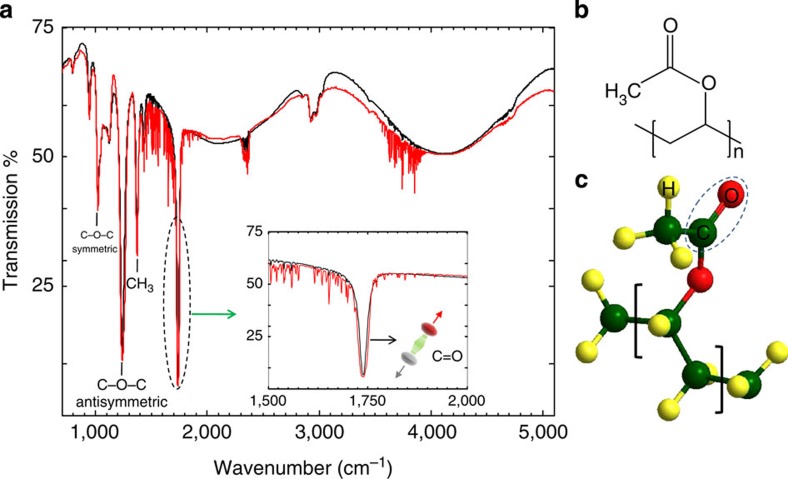
    1. Bower D. I. & Maddams W. F. Vibrational Spectroscopy of Polymers Cambridge Univ. Press (1992).
    1. Voltz R. Theory of molecular decay processes. Org. Mol. Photophys. 2, 217–302 (1975).
    1. Barron L. Molecular Light Scattering and Optical Activity 2nd edn Cambridge Univ. Press (2004).
    1. Houdré R., Stanley R. P. & Ilegems M. Vacuum-field Rabi splitting in the presence of inhomogeneous broadening: Resolution of a homogeneous linewidth in an inhomogeneously broadened system. Phys. Rev. A 53, 2711–2715 (1996). - PubMed
    1. Agranovich V. M., Gartstein Y. N. & Litinskaya M. Hybrid resonant organic-inorganic nanostructures for optoelectronic applications. Chem. Rev. 111, 5179–5214 (2011). - PubMed

Publication types

LinkOut - more resources