Review
. 2015 Mar;38(3):158-66.
doi: 10.1016/j.tins.2014.12.007. Epub 2015 Jan 9.

Amygdala-prefrontal interactions in (mal)adaptive learning

Affiliations
Review

Amygdala-prefrontal interactions in (mal)adaptive learning

Ekaterina Likhtik et al. Trends Neurosci. 2015 Mar.

Abstract

The study of neurobiological mechanisms underlying anxiety disorders has been shaped by learning models that frame anxiety as maladaptive learning. Pavlovian conditioning and extinction are particularly influential in defining learning stages that can account for symptoms of anxiety disorders. Recently, dynamic and task related communication between the basolateral complex of the amygdala (BLA) and the medial prefrontal cortex (mPFC) has emerged as a crucial aspect of successful evaluation of threat and safety. Ongoing patterns of neural signaling within the mPFC-BLA circuit during encoding, expression and extinction of adaptive learning are reviewed. The mechanisms whereby deficient mPFC-BLA interactions can lead to generalized fear and anxiety are discussed in learned and innate anxiety. Findings with cross-species validity are emphasized.

Keywords: amygdala; generalization; oscillations; pavlovian learning; prefrontal cortex; safety.


Figures

Figure 1
Figure 1
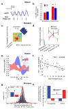
mPFC – BLA engagement dynamically changes during training and recall of aversive learning. (A–B) Neurons in the BLA are synchronized with theta oscillations (4–12 Hz) in the mPFC during stimulus assessment (mouse). (A) Example of a simultaneously recorded local field potential (LFP) oscillation (black – raw trace; blue –theta filtered trace) and firing of a single unit. (B) Mice that discriminate between the CS+ and CS− during recall, show increased mPFC-BLA synchrony. In mice that generalize fear to both stimuli, mPFC-BLA synchrony does not increase above baseline during stimulus presentation. MRL- mean resultant length. (C–D) mPFC-BLA synchrony is higher during stimulus assessment (monkey). (C) Example of simultaneously recorded neural firing in the dACC and BLA during the end of training on a partial reinforcement schedule. After CS onset, neural firing in the two areas is highly correlated. (D) Whereas correlated neural firing in dACC-BLA remains high throughout training when monkeys are trained on a partial reinforcement schedule (red trace), correlated firing drops quickly during training on a continuous reinforcement schedule (green trace). (E–F) During discrimination of a safe CS−, BLA firing is predominantly coupled to prefrontal theta oscillations of the past. (E) BLA cells of mice that discriminate between an aversive CS+ and a safe CS− are more synchronized (phase-locked) to mPFC theta oscillations of the past only during the safe CS− (blue). During the aversive CS+ (red) and both the CS+ and CS− of mice that generalize fear (inset), there is no preferred direction of information flow between the BLA and mPFC. (F) Higher probability of prefrontal theta oscillations leading changes in amygdala theta oscillations during the CS− correlates with better discriminated between the two stimuli (Discrimination Score). (G–H) Dynamic changes in BLA-dACC communication during adaptive learning in the macaque. (G) Histogram of the center-of-masses (CoMs) obtained from all significant cross-correlations of simultaneously recorded spikes in the BLA and dACC during learning. Cross-correlations of spiking activity shows that during learning (red) communication between the two regions increases and becomes more bidirectional than during the habituation period, when the dACC tends to lead the amygdala (blue). The variance of the distribution is significantly smaller during learning than habituation (Inset), indicating a tighter coupling of activity between the two areas. (H) Neurons in the amygdala fire before dACC (blue) when an amygdala cell fires to all stimuli, (ones that predict an upcoming stimulus and ones that do not). However, dACC neurons lead amygdala neural firing in cells that fire in response to stimuli that don’t deliver an expected outcome. Figures adapted with authors’ permission [57,48,60].
Figure 2
Figure 2
Cross-species findings in mPFC-BLA circuit function during adaptive learning. Similar findings in more than one species are highlighted by boxes. The colored dots refer to the techniques used to obtain the described findings. Technique Key is found in the upper left. Images of brains are published with permission, courtesy of the University of Wisconsin and Michigan State Comparative Mammalian Brain Collections, and the National Museum of Health and Medicine (brainmuseum.org). All preparation of the specimens and images were funded by the NSF. Rat, Mouse, Primate silhouettes are Wikimedia images in the public domain. Human figure, drawing courtesy of P. Drubetskoy.

References

    1. Pavlov I. Conditioned reflexes: an investigation of the physiological activity of the cerebral cortex. London: Oxford University Press; 1927. - PMC - PubMed
    1. Poulos AM, Fanselow MS. The neuroscience of mammalian associative learning. Annu Rev Psychol. 2005;56:207–234. - PubMed
    1. Pabba M. Evolutionary development of the amygdaloid complex. Front Neuroanat. 2013;2:27. - PMC - PubMed
    1. Medina L, Bupesh M, Abellán A. Contribution of genorarchitecture to understanding forebrain evolution and development, with a particular emphasis on the amygdala. Brain Behav Evol. 2011;78:216–236. - PubMed
    1. McDonald AJ. Cortical pathways to the mammalian amygdala. Prog Neurobiol. 1998;55:257–332. - PubMed

Publication types