Clinical Trial
. 2015 Jan 27;112(4):1208-13.
doi: 10.1073/pnas.1412389112. Epub 2015 Jan 12.

New human-specific brain landmark: the depth asymmetry of superior temporal sulcus

Affiliations
Clinical Trial

New human-specific brain landmark: the depth asymmetry of superior temporal sulcus

François Leroy et al. Proc Natl Acad Sci U S A. .

Abstract

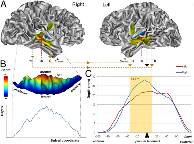
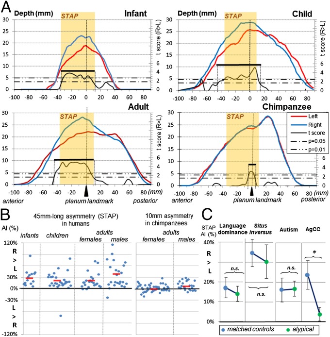
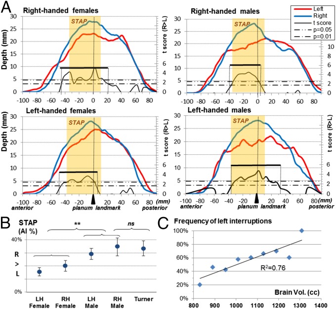
Identifying potentially unique features of the human cerebral cortex is a first step to understanding how evolution has shaped the brain in our species. By analyzing MR images obtained from 177 humans and 73 chimpanzees, we observed a human-specific asymmetry in the superior temporal sulcus at the heart of the communication regions and which we have named the "superior temporal asymmetrical pit" (STAP). This 45-mm-long segment ventral to Heschl's gyrus is deeper in the right hemisphere than in the left in 95% of typical human subjects, from infanthood till adulthood, and is present, irrespective of handedness, language lateralization, and sex although it is greater in males than in females. The STAP also is seen in several groups of atypical subjects including persons with situs inversus, autistic spectrum disorder, Turner syndrome, and corpus callosum agenesis. It is explained in part by the larger number of sulcal interruptions in the left than in the right hemisphere. Its early presence in the infants of this study as well as in fetuses and premature infants suggests a strong genetic influence. Because this asymmetry is barely visible in chimpanzees, we recommend the STAP region during midgestation as an important phenotype to investigate asymmetrical variations of gene expression among the primate lineage. This genetic target may provide important insights regarding the evolution of the crucial cognitive abilities sustained by this sulcus in our species, namely communication and social cognition.

Keywords: STS; anatomy; asymmetry; brain; human-specific.


Conflict of interest statement

The authors declare no conflict of interest.

Figures

Fig. 1.
Fig. 1.
(A) Location of the STAP (yellow) relative to Heschl’s gyrus (blue) and the ventral tip of the central sulcus (green) on both left and right inner cortical surfaces of an individual adult brain. The STAP center is shown by a cross. The black dot with a white contour line shows the planum temporale landmark. (B, Upper) Sulcal depth shown by color coding of the sulcal mesh (seen from above). (Lower) Sulcal depth profile in the right hemisphere of an individual subject. (C) Adult sulcal depth profile; STAP anterior and posterior ends as well as the planum landmark are drawn in dotted lines. The light orange overlay illustrates the STAP (deeper on the right), defined as the common asymmetrical segment in the three typical groups (infant, right-handed children, and adults).
Fig. 2.
Fig. 2.
(A) Left (red line) and right (blue line) STS depth profiles from the temporal pole to its parietal caudal end. Depth profiles are shown for infants, right-handed children, adults, and chimpanzees. The asymmetrical part of the STS is computed for each group by permutation tests over 5-mm-long intervals along the sulcus. Two statistical thresholds (Pcorr = 0.05; Pcorr = 0.01) are shown by horizontal dashed-and-dotted lines. The extent of the asymmetrical segment is given by the range of the Student t variable (black line) above the lower threshold and is identified by a black bar (see also Table 1). The extent of the common region across the three typical human groups (STAP) is shown in light orange overlay. (B) STAP per cent in individual typical humans and chimpanzees. Mean group values are shown by a short red line. (C) STAP in atypical human groups, i.e., adults with right lateralization for language, adults with situs inversus (reversed petalia), children with autism spectrum disorders, and children with AgCC. *P < 0.05. n.s., not significant.
Fig. 3.
Fig. 3.
The STAP in relation to sex and handedness in adults. (A) Left (red line) and right (blue line) STS depth profiles. The extent of the asymmetrical segment is given by the range of the Student t variable (black line) above the lower threshold and is identified by a black bar (see also Table 1). The extent of the STAP region is shown in light orange overlay. (B) STAP statistics (mean and SE) in healthy right-handers and left-handers and in patients with Turner syndrome. (C) Frequency of left sulcal interruptions in relation to brain volume. LH, left-handed; n.s., not significant; RH, right-handed. **P < 0.01.

References

    1. Geschwind N, Levitsky W. Human brain: Left-right asymmetries in temporal speech region. Science. 1968;161(3837):186–187. - PubMed
    1. Dorsaint-Pierre R, et al. Asymmetries of the planum temporale and Heschl’s gyrus: Relationship to language lateralization. Brain. 2006;129(Pt 5):1164–1176. - PubMed
    1. Keller SS, Crow T, Foundas A, Amunts K, Roberts N. Broca’s area: Nomenclature, anatomy, typology and asymmetry. Brain Lang. 2009;109(1):29–48. - PubMed
    1. Hopkins WD, Nir TM. Planum temporale surface area and grey matter asymmetries in chimpanzees (Pan troglodytes): The effect of handedness and comparison with findings in humans. Behav Brain Res. 2010;208(2):436–443. - PMC - PubMed
    1. Pelphrey KA, Viola RJ, McCarthy G. When strangers pass: Processing of mutual and averted social gaze in the superior temporal sulcus. Psychol Sci. 2004;15(9):598–603. - PubMed

Publication types

MeSH terms