. 2015 Jan 27;112(4):1047-52.
doi: 10.1073/pnas.1411610112. Epub 2015 Jan 12.

Visualization of the type III secretion sorting platform of Shigella flexneri

Affiliations

Visualization of the type III secretion sorting platform of Shigella flexneri

Bo Hu et al. Proc Natl Acad Sci U S A. .

Abstract

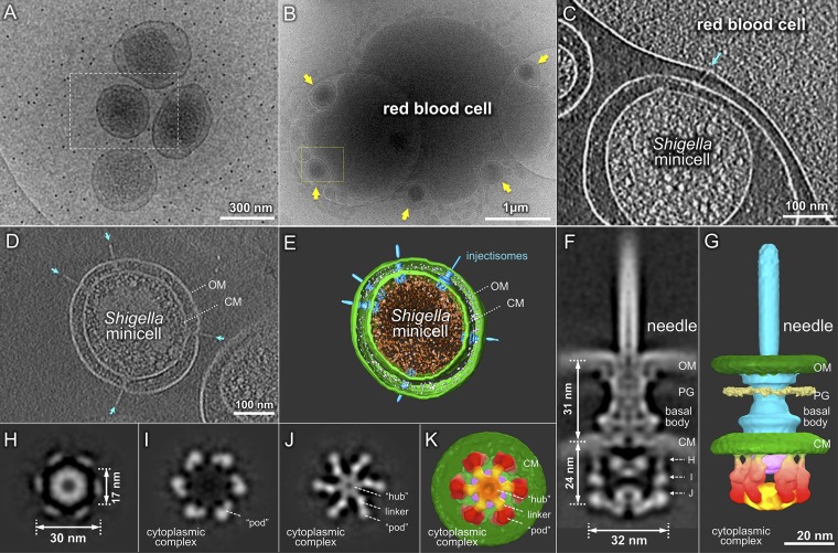
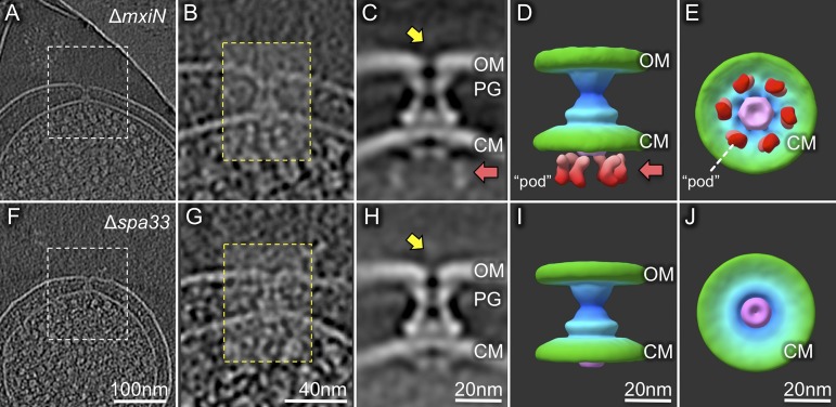
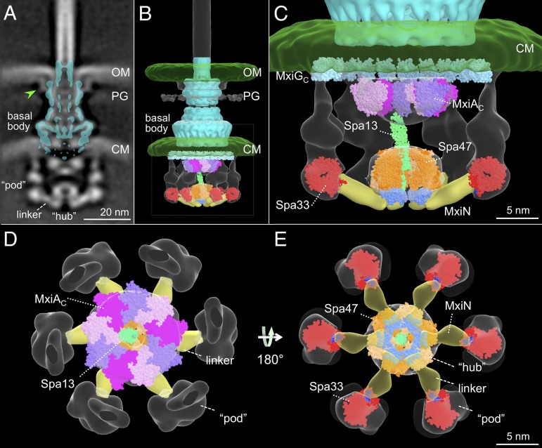
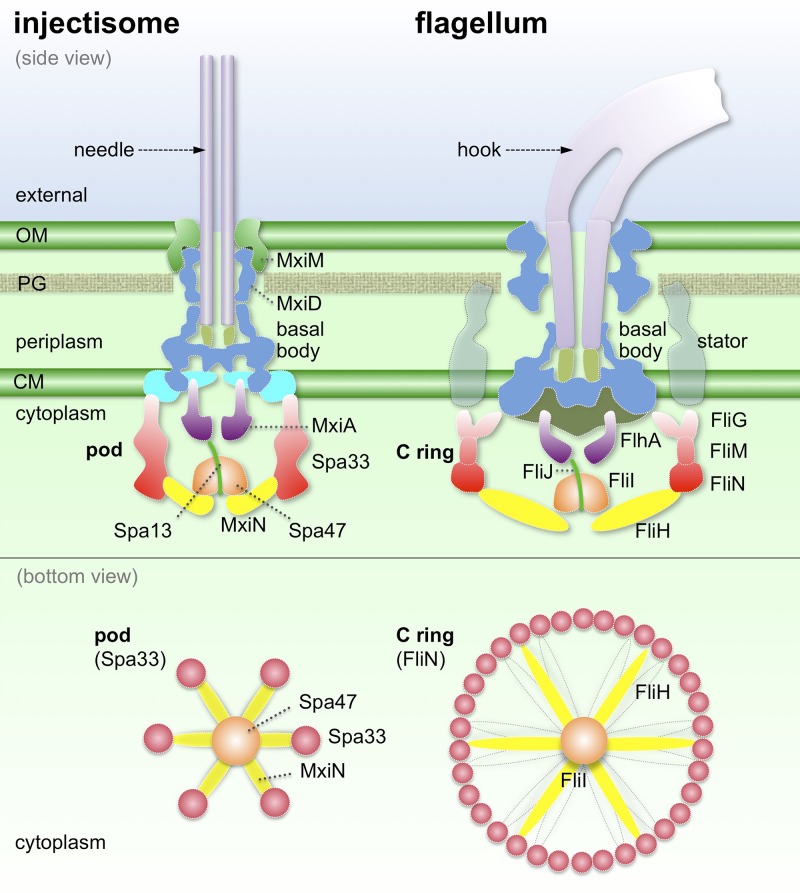
Bacterial type III secretion machines are widely used to inject virulence proteins into eukaryotic host cells. These secretion machines are evolutionarily related to bacterial flagella and consist of a large cytoplasmic complex, a transmembrane basal body, and an extracellular needle. The cytoplasmic complex forms a sorting platform essential for effector selection and needle assembly, but it remains largely uncharacterized. Here we use high-throughput cryoelectron tomography (cryo-ET) to visualize intact machines in a virulent Shigella flexneri strain genetically modified to produce minicells capable of interaction with host cells. A high-resolution in situ structure of the intact machine determined by subtomogram averaging reveals the cytoplasmic sorting platform, which consists of a central hub and six spokes, with a pod-like structure at the terminus of each spoke. Molecular modeling of wild-type and mutant machines allowed us to propose a model of the sorting platform in which the hub consists mainly of a hexamer of the Spa47 ATPase, whereas the MxiN protein comprises the spokes and the Spa33 protein forms the pods. Multiple contacts among those components are essential to align the Spa47 ATPase with the central channel of the MxiA protein export gate to form a unique nanomachine. The molecular architecture of the Shigella type III secretion machine and its sorting platform provide the structural foundation for further dissecting the mechanisms underlying type III secretion and pathogenesis and also highlight the major structural distinctions from bacterial flagella.

Keywords: cryo-electron tomography; injectisome; nanomachine; pathogen–host interaction; protein secretion.


Conflict of interest statement

The authors declare no conflict of interest.

Figures

Fig. 1.
Fig. 1.
Cryo-ET of S. flexneri minicells reveals intact T3SS and its cytoplasmic structure. (A) A cryo-EM image shows tiny S. flexneri minicells with diameters of ∼0.3 μm. Purified S. flexneri minicells are able to interact intimately with an RBC (B). Yellow arrows highlight the five minicells adhering to the RBC, whose membrane is indented at each contact point. A tomographic slice reveals that an injectisome (indicated by a cyan arrow) is directly involved in the interaction with the host cell membrane (C). (D) A central slice and (E) a 3D surface rendering of a tomographic reconstruction of a typical minicell show multiple injectisomes embedded in the cell envelope, including outer membrane (OM) and cytoplasmic membrane (CM). A central section (F) and a 3D surface rendering (G) of the subtomogram average of the intact injectisome show OM, CM, peptidoglycan (PG), basal body, and needle in detail. Importantly, there is a large cytoplasmic complex that is 32 nm in diameter and 24 nm in height (F). Three cross-sections (indicated in F) of the cytoplasmic complex show sixfold symmetric features (H–J). The bottom view (K) and a side view (G) of the injectisome present the apparent discontinuity of the outer ring of six “pod-like” densities. The six pods (colored in red) are linked to the central hub (orange) by radially arranged (spoke-like) linker densities (yellow).
Fig. 2.
Fig. 2.
Injectisome structural differences in S. flexneri minicell mutants lacking either MxiN or Spa33. Analysis of ∆mxiN (A–E) or ∆spa33 (F–J) minicells is shown. Depicted are representative slices of cryo-ET reconstructions of a ∆mxiN minicell (A) or a ∆spa33 minicell (F), followed by the corresponding zoomed-in views (B and G), averaged structures (C and H), and two 3D surface renderings (D, E, and I–J). Both mutants lack the needle (yellow arrows) and the central hub of the cytoplasmic domain (Fig. 1 G and J). The ∆spa33 injectisomes also lack the six outer densities (pods) of the cytoplasmic domain (red arrows and red-colored densities seen in the ∆mxiN injectisomes). The predicted location of the MxiA complex is indicated in purple in the surface renderings.
Fig. 3.
Fig. 3.
A molecular model of the T3SS injectisome. The isolated Shigella T3SS basal body (10) is fitted onto the intact injectisome map in a central section (A) and a surface rendering (B). Extra densities are apparent, including outer membrane (OM), cytoplasmic membrane (CM), peptidoglycan (PG), a large cytoplasmic complex, and a base element (green arrowhead) where it interacts with the OM. Models of several cytoplasmic proteins (MxiAC, Spa47, Spa13, Spa33) are fitted in the surface rendering map. Zoomed-in views of the cytoplasmic portion of B are shown from the side (C), top (D), and bottom (E). The Spa47 ATPase hexameric ring (the homolog of V-ATPase, PDB ID code 3J0J, orange) together with Spa13 (the homolog of FliJ, PDB ID code 3AJW, green) is docked into the central hub of the map. Spa13 is long enough to interact with the nonameric ring of MxiAC (different shades of purple), which fits well into the torus-like structure near the cytoplasmic membrane (C and D). Underneath the MxiGC ring (the homolog of PrgHC, PDB ID code 3J1W, dark green), six spa33-encoded complexes form the proposed sorting platform. We place the Spa33 homologs (FliN tetramer, PDB ID code 1YAB, red) into the bottom part of the pod. MxiN forms the spoke-like linker (yellow) that interacts with both the hydrophobic patch (blue) in Spa33 and the C-terminal domain (cyan) of Spa47 as shown in the bottom view (E).
Fig. 4.
Fig. 4.
Comparative structural models of injectisomes and bacterial flagella. The cytoplasmic hexamer of the Spa33 complex positions the Spa47 ATPase (via MxiN) directly beneath and in line with the export gate, beginning with the MxiA cytoplasmic domain. In contrast, the flagellar C ring contains multiple copies of FliG, FliM, and FliN that are involved in secretion, rotation, and switching. FliH, the homolog of MxiN, is likely sufficiently flexible to interact with different FliN proteins at the bottom of the C ring. The outer membrane (OM), cytoplasmic membrane (CM), peptidoglycan (PG), a large cytoplasmic complex, and the basal body are colored consistently as those in previous figures.

References

    1. Cornelis GR. The type III secretion injectisome. Nat Rev Microbiol. 2006;4(11):811–825. - PubMed
    1. Galán JE, Wolf-Watz H. Protein delivery into eukaryotic cells by type III secretion machines. Nature. 2006;444(7119):567–573. - PubMed
    1. Kubori T, et al. Supramolecular structure of the Salmonella typhimurium type III protein secretion system. Science. 1998;280(5363):602–605. - PubMed
    1. Deane JE, Abrusci P, Johnson S, Lea SM. Timing is everything: The regulation of type III secretion. Cell Mol Life Sci. 2010;67(7):1065–1075. - PMC - PubMed
    1. Lara-Tejero M, Kato J, Wagner S, Liu X, Galán JE. A sorting platform determines the order of protein secretion in bacterial type III systems. Science. 2011;331(6021):1188–1191. - PMC - PubMed

Publication types

Substances

LinkOut - more resources