. 2015 Mar;83(3):1181-9.
doi: 10.1128/IAI.02788-14. Epub 2015 Jan 12.

Importance of bacterial replication and alveolar macrophage-independent clearance mechanisms during early lung infection with Streptococcus pneumoniae

Affiliations

Importance of bacterial replication and alveolar macrophage-independent clearance mechanisms during early lung infection with Streptococcus pneumoniae

Emilie Camberlein et al. Infect Immun. 2015 Mar.

Abstract

Although the importance of alveolar macrophages for host immunity during early Streptococcus pneumoniae lung infection is well established, the contribution and relative importance of other innate immunity mechanisms and of bacterial factors are less clear. We have used a murine model of S. pneumoniae early lung infection with wild-type, unencapsulated, and para-amino benzoic acid auxotroph mutant TIGR4 strains to assess the effects of inoculum size, bacterial replication, capsule, and alveolar macrophage-dependent and -independent clearance mechanisms on bacterial persistence within the lungs. Alveolar macrophage-dependent and -independent (calculated indirectly) clearance half-lives and bacterial replication doubling times were estimated using a mathematical model. In this model, after infection with a high-dose inoculum of encapsulated S. pneumoniae, alveolar macrophage-independent clearance mechanisms were dominant, with a clearance half-life of 24 min compared to 135 min for alveolar macrophage-dependent clearance. In addition, after a high-dose inoculum, successful lung infection required rapid bacterial replication, with an estimated S. pneumoniae doubling time of 16 min. The capsule had wide effects on early lung clearance mechanisms, with reduced half-lives of 14 min for alveolar macrophage-independent and 31 min for alveolar macrophage-dependent clearance of unencapsulated bacteria. In contrast, with a lower-dose inoculum, the bacterial doubling time increased to 56 min and the S. pneumoniae alveolar macrophage-dependent clearance half-life improved to 42 min and was largely unaffected by the capsule. These data demonstrate the large effects of bacterial factors (inoculum size, the capsule, and rapid replication) and alveolar macrophage-independent clearance mechanisms during early lung infection with S. pneumoniae.


Figures

FIG 1
FIG 1
The capsule inhibits AM phagocytosis of S. pneumoniae during early lung infection. (A and B) Association of 6-carboxyfluorescein–succinimidyl ester (FAM-SE)-labeled S. pneumoniae with AMs recovered from mice 4 h after infection by i.n. inoculation with 5 × 10⁶ CFU of TIGR4 (white boxes) or P1672 (gray boxes) assessed by flow cytometry and presented as percentage of fluorescent AMs (A) and the geometric mean fluorescence intensity of fluorescent AMs (B) (14). Representative of data obtained from repeated experiments. (C) Confocal assessment of the internalization of FAM-SE-labeled S. pneumoniae by AMs recovered from mice 4 h after i.n. inoculation with 5 × 10⁶ CFU of TIGR4 or P1672. The data show the median (and interquartile range [IQR]) numbers of intracellular bacteria identified by Z stacking for AMs containing S. pneumoniae. P values were obtained using Mann-Whitney U tests. (D) Representative examples of confocal microscopy of recovered AMs. Blue, DAPI staining of the nuclei; red, F4/80 staining (macrophage marker); green, FAM-SE-labeled bacteria.
FIG 2
FIG 2
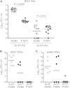
Effects of inoculum size and the capsule on S. pneumoniae BALF CFU during early lung infection. (A) Median BALF CFU (log10/ml) 4 h after i.n. inoculation with 5 × 10⁶ or 5 × 10⁵ CFU of TIGR4 (encapsulated; white circles) and P1672 (unencapsulated; gray symbols) S. pneumoniae. Pooled data are from two separate experiments and representative of repeated experiments. BALF (B) and spleen (C) CFU 24 h after i.n. inoculation with 5 × 10⁶ (white symbols) or 5 × 10⁵ (black symbols) CFU of TIGR4 or 5 × 10⁶ CFU of P1672 (gray symbols) S. pneumoniae. Each symbol represents results for an individual mouse, and bars represent median CFU/ml. P values were obtained using Mann-Whitney U tests.
FIG 3
FIG 3
Effects of macrophage depletion on S. pneumoniae CFU in BALF during early lung infection. (A and B) Median BALF CFU (log10/ml) 4 h after i.n. inoculation with 5 × 10⁶ (A) or 5 × 10⁵ (B) CFU of TIGR4 or P1672 S. pneumoniae in mice pretreated with control liposomes (AM+; white symbols) or clodronate liposomes (AM−; black symbols). Each symbol represents results for an individual mouse, bars represents median CFU/ml, and P values were obtained using Mann-Whitney U tests.
FIG 4
FIG 4
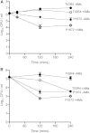
Time course of BALF CFU over the initial 4 h of S. pneumoniae lung infection used for mathematical modeling of AM-dependent clearance half-lives. (A) BALF CFU 30, 120, and 240 min after i.n. inoculation with 5 × 10⁶ (log10 6.7) CFU of TIGR4 (circles) or P1672 (diamonds) S. pneumoniae in mice pretreated with control liposomes (white symbols; +AMs) or clodronate liposomes (black symbols; −AMs). Data were pooled from two experiments with 9 to 14 mice each. (B) BALF CFU 120 and 240 min after i.n. inoculation with 5 × 10⁵ (log10 5.7) CFU of TIGR4 (circles) or P1672 (diamonds) S. pneumoniae in mice pretreated with control liposomes (white symbols; +AMs) or clodronate liposomes (black symbols; −AMs). Data were from one experiment that was representative of two different experiments with 6 mice each. The data were plotted assuming a starting CFU within BALF of log10 6.2 (A) or 5.2 (B); symbols represent mean BALF CFU/ml, and error bars represent standard deviations (SDs) (where not shown error bars are contained within symbols).
FIG 5
FIG 5
Mean (SD) BALF neutrophilia 30, 120, and 240 min after inoculation with 5 × 10⁶ CFU of TIGR4 (gray columns) or P1672 (white columns) S. pneumoniae in mice pretreated with control liposomes (solid columns) or clodronate liposomes (hatched columns). There were no significant differences between groups at each time point (Mann-Whitney U tests). Data are from one experiment that was representative of repeated experiments with 6 mice each.
FIG 6
FIG 6
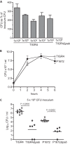
Effects of bacterial replication on S. pneumoniae BALF CFU for growth in BALF in vitro and during early lung infection. (A) Growth in 1 ml of BALF in vitro of different inoculum sizes of TIGR4 (stated beneath each column) and 1 × 10⁶ CFU TIGR4Δpab expressed as a percentage of the initial inoculum. Error bars represent standard errors of the means (SEMs). The results for TIGR4Δpab were <1%. (B) Growth in BALF in vitro of S. pneumoniae TIGR4 (white symbols) or P1672 (black symbols) expressed as mean (SEM) CFU/ml over time. (C) Median BALF CFU (log10/ml) 4 h after i.n. inoculation with 5 × 10⁶ of TIGR4 (white symbols), TIGR4Δpab (gray symbols), P1672 (white symbols), or P1672Δpab (gray symbols). Each symbol represents results for an individual mouse, bars represents median CFU/ml, and P values were obtained using Mann-Whitney U tests.
FIG 7
FIG 7
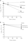
BALF CFU data used for calculation of S. pneumoniae replication times during early lung infection. The data show the numbers of BALF CFU 30 and 120 min after i.n. inoculation with 5 × 10⁶ (log10 6.7) CFU (A) or 120 min after i.n. inoculation with 5 × 10⁵ (log10 5.7) (B) of TIGR4, TIGR4Δpab, P1672, or P1672Δpab. The data are representative of repeated experiments and plotted assuming a starting CFU in BALF of log10 6.2 (A) or 5.2 (B). Symbols represent mean BALF CFU/ml, error bars represent SDs (where not shown, error bars are contained within symbols), and n = 6 to 10.

References

    1. Gordon SB, Read RC. 2002. Macrophage defences against respiratory tract infections. Br Med Bull 61:45–61. doi:10.1093/bmb/61.1.45. - DOI - PubMed
    1. Green GM, Kass E. 1964. The role of the alveolar macrophage in the clearance of bacteria from the lung. J Exp Med 119:167–176. doi:10.1084/jem.119.1.167. - DOI - PMC - PubMed
    1. Kim M, Goldstein E, Lewis JP, Lippert W, Warshauer D. 1976. Murine pulmonary alveolar macrophages: rates of bacterial ingestion, inactivation, and destruction. J Infect Dis 133:310–320. doi:10.1093/infdis/133.3.310. - DOI - PubMed
    1. Lee H-Y, Andalibi A, Webster P, Moon S-K, Teufert K, Kang S-H, Li J-D, Nagura M, Ganz T, Lim DJ. 2004. Antimicrobial activity of innate immune molecules against Streptococcus pneumoniae, Moraxella catarrhalis and nontypeable Haemophilus influenzae. BMC Infect Dis 4:12. doi:10.1186/1471-2334-4-12. - DOI - PMC - PubMed
    1. Rogan MP, Geraghty P, Greene CM, O'Neill SJ, Taggart CC, McElvaney NG. 2006. Antimicrobial proteins and polypeptides in pulmonary innate defence. Respir Res 7:29. doi:10.1186/1465-9921-7-29. - DOI - PMC - PubMed

Publication types

MeSH terms

Substances

LinkOut - more resources