. 2015 Feb 9;212(2):149-63.
doi: 10.1084/jem.20141505. Epub 2015 Jan 12.

Bee venom processes human skin lipids for presentation by CD1a

Affiliations

Bee venom processes human skin lipids for presentation by CD1a

Elvire A Bourgeois et al. J Exp Med. .

Abstract

Venoms frequently co-opt host immune responses, so study of their mode of action can provide insight into novel inflammatory pathways. Using bee and wasp venom responses as a model system, we investigated whether venoms contain CD1-presented antigens. Here, we show that venoms activate human T cells via CD1a proteins. Whereas CD1 proteins typically present lipids, chromatographic separation of venoms unexpectedly showed that stimulatory factors partition into protein-containing fractions. This finding was explained by demonstrating that bee venom-derived phospholipase A2 (PLA2) activates T cells through generation of small neoantigens, such as free fatty acids and lysophospholipids, from common phosphodiacylglycerides. Patient studies showed that injected PLA2 generates lysophospholipids within human skin in vivo, and polyclonal T cell responses are dependent on CD1a protein and PLA2. These findings support a previously unknown skin immune response based on T cell recognition of CD1a proteins and lipid neoantigen generated in vivo by phospholipases. The findings have implications for skin barrier sensing by T cells and mechanisms underlying phospholipase-dependent inflammatory skin disease.


Figures

Figure 1.
Figure 1.
Vespula spp and Apis mellifera venoms induce a preferential activation of CD1a-restricted T cells among the group1-CD1 reactive cells. (A) T cells were isolated by CD3 MACS beads from healthy donor PBMCs and cultured for 12–14 d with IL-2 and irradiated K562 cells transfected with CD1a (K562-CD1a), CD1b (K562-CD1b), CD1c (K562-CD1c), CD1d (K562-CD1d), or an empty vector (K562) in the presence of venom. CD1 reactivity was then examined by IFN-γ ELISpot with transfected or untransfected K562 cells either in the presence or absence of Vespula spp or Apis mellifera venoms. Representative data for one donor (C1175) of three are shown. (B) The CD1a-restricted T cell response in the presence or absence of anti-CD1a (donor C1098) and Vespula spp and Apis mellifera venoms. CD1a-restricted, venom-specific responses were measured in 21 donors for Vespula spp venom (C) and Apis mellifera venom (D). mDC or in vitro LC–like cells derived from CD14+ cells were pulsed with 1 µg/ml wasp venom (E) or bee venom (F) and incubated with the T cells in the presence or absence of anti-CD1a antibody or isotype control. IFN-γ production was measured by IFN-γ ELISpot. Representative data for one donor (C556 [E] and C560 [F]) of three are shown. Data were mean of triplicate measurements ± SEM. *, P < 0.05; **, P < 0.01; ***, P < 0.001.
Figure 2.
Figure 2.
The autoreactive CD1a-restricted T cell clone BC2 is activated by wasp and bee venoms. Biotinylated CD1a, CD1b, and CD1c and CD1d proteins were coated on streptavidin plates. After washing, wasp (A and B) and bee (C) venoms and dideoxymycobactin (DDM; B) were added to the wells for 24–48 h. After washing, BC2 cells (A and C) or CD8.2 cells (B) were added, and the supernatants were collected 24 h later and IFN-γ quantified. (D) BC2 cells were co-cultured for 24 h with universal target cells (K562) as APCs, transfected with CD1a (K562-CD1a) or empty vector (K562) and bee venom. IFN-γ in supernatants was measured by ELISA. Data are representative of three separate experiments. (E) mDCs and (F) CD34-derived LC-like cells were incubated with IgG control or anti-CD1a antibodies for 1 h at 37°C. Bee venom was then added at 2 µg/ml for 1 h. BC2 cells were then added at a 1:25 APC/T cell ratio. Two representative donors out of three are shown.
Figure 3.
Figure 3.
The co-incubation of both proteins and lipids from wasp and bee venoms triggers BC2 cells activation when presented by CD1a protein. (A) Protein and lipid fractions of bee and wasp venoms were separated by organic solvent extraction using chloroform and methanol. (B) CD1a proteins were coated on streptavidin plates. After washing, wasp or bee venoms and their extracts were added to the wells for 24 to 48 h. After washing, BC2 cells were added, supernatants were collected after 24 h and IFN-γ quantified. (C) BC2 cells were co-cultured with universal target cells transfected with CD1a (K562 CD1a) or empty vector (K562) and bee venom or their extracts. Supernatants were collected after 24 h. IFN-γ in supernatants was measured by ELISA. Data are representative of three separate experiments.
Figure 4.
Figure 4.
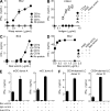
Bee venom PLA2 alone induces a CD1a-restricted T cell response by releasing free fatty acids. BC2 cells were co-cultured for 24 h with universal target cells (K562) transfected with CD1a (K562 CD1a) or empty vector (K562) and bee venom or bee venom PLA2 (A), mastoparan (B), or arachidonic acid (C). IFN-γ in supernatants was measured by ELISA. In (C) right panel, biotinylated CD1a proteins were coated on streptavidin plates. After washing, bee venom or arachidonic acid were added to the wells for 24 to 48 h. After washing, BC2 cells were added for 24 h, and supernatant was sampled for IFN-γ quantification by ELISA. Data are representative of three separate experiments. (D) mDC and (E) CD34-derived LC-like cells were incubated with IgG control or anti-CD1a antibodies for 1 h at 37C. PLA2 was then added at 2 µg/ml for 1 h. BC2 cells were then added at a 1:25 APC:T cell ratio. Two representative donors out of three are shown.
Figure 5.
Figure 5.
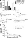
Wasp venom phospholipase is active in vivo in the skin. (A) Biotinylated CD1a proteins were coated on streptavidin plates. After washing, lipids were added at 10 µg/ml for 24–48 h. Lipids tested were as follows: phosphatidylcholine 18:1 (PC18:1), lysophosphatidylcholine 18:1 (LPC18:1), and oleic acid 18:1 (FA 18:1; left) or phosphatidic acid 16:0 (PA16:0), lysophosphatidic acid 18:1 (LPA18:1), and palmitic acid 16:0 (FA16:0; right). Phosphatidylcholine and lysophosphatidic acid were preincubated or not with PLA2 at 50 µg/ml for 1 h at 37°C. 24 h later, wells were washed and BC2 cells were added. Supernatants were collected after 24 h. IFN-γ in supernatants was measured by ELISA. Data are representative of three separate experiments. (B) Healthy donor epidermis was injected with 10 µg of wasp venom or saline. Skin blisters were raised and, 24 h later, the blister fluid was sampled. Lipids were extracted with chloroform, methanol, and water using the Bligh-Dyer method and analyzed on a quadruple time of flight mass spectrometer. Positive mode EIC-MS detected ions at m/z 758.5686, 786.6000, 810.5994, and 834.5993, corresponding to the homologous series of phosphatidylcholine C42H81NO8P+ (16:0/18:2), C44H85NO8P+ (18:0/18:2), C46H85NO8P+ (18:2/20:2), and C48H85NO8P+ (20:4/20:2). Positive mode EIC-MS detected ions at m/z 496.3374, 524.3683, and 544.3370, corresponding to the homologous series of lysophosphatidylcholine C24H51NO7P+ (16:0), C26H55NO7P+ (18:0), and C28H51NO7P+ (20:4). Data from one representative donor out of two are shown.
Figure 6.
Figure 6.
Vespula spp and Apis mellifera venom PLA2 activity. (A) PLA2 activity was detected as free thiol groups released from thiol-labeled lipid substrates through addition of DNTB/EGTA and measuring color generation at 415 nm using Apis mellifera venom with four phosphatidylcholine-based substrates of varying acyl chain lengths and saturation, but the same headgroup. Vespula spp venom PLA2 activity for substrates diheptanoyl-phosphatidylcholine and arachidonyl-phosphatidylcholine (P < 0.001 compared with no enzyme control). (B) Using lipid substrate with a thiol group at the sn2 position, PLA2 activity in Vespula spp venom in the presence or absence of manoalide was detected using DNTB/EGTA, which produces a colored precipitate and measured at 415 nm. Data were mean of triplicate measurements ± SEM.
Figure 7.
Figure 7.
CD1a-restricted reactivity to Apis mellifera venom PLA2 is found among polyclonal T cells from blood of healthy donors. CD3+ cells were isolated by magnetic beads from healthy donor PBMC and cultured for 12–14 d with IL-2 and irradiated K562 cells transfected with CD1a (K562 CD1a) or CD1b (K562 CD1b) or CD1c (K562 CD1c) or CD1d (K562 CD1d) or an empty vector (K562) in the presence of Apis mellifera venom PLA2. CD1 reactivity was then examined by IFN-γ ELISpot with transfected or untransfected K562 cells either in the presence or absence of Apis mellifera venom 100 ng/ml PLA2 (A, left). Data from one representative donor of three are shown. Venom PLA2 specific CD1a-restricted T cell responses were measured in 18 donors (A, right). CD1a-restricted responses were examined in the presence or absence of anti-CD1a (B, left) for donor C1098, and PLA2-neutralizing antibodies (7B, right), and manoalide (specific PLA2 inhibitor) for wasp venom in donor C559 (C, left) and Apis mellifera venom PLA2 in donor C334 (C, right). Representative results are shown from three independent experiments. (D) IFN-γ production was measured by co-incubations of PLA2-specific T cells with mDCs (left) or CD14-derived LC-like DCs (middle) in the presence or absence of anti-CD1a for donor C558. IFN-γ production was measured from co-incubations of PLA2-specific T cells and freshly isolated skin CD1a+ cells in the presence or absence of anti-CD1a or manoalide (right). Representative data from 3 (left and middle) or 2 (right) separate donors are shown. Data are mean of triplicate measurements ± SEM. *, P < 0.05; **, P < 0.01; ***, P < 0.001.

Comment in

References

    1. Agea E., Russano A., Bistoni O., Mannucci R., Nicoletti I., Corazzi L., Postle A.D., De Libero G., Porcelli S.A., and Spinozzi F.. 2005. Human CD1-restricted T cell recognition of lipids from pollens. J. Exp. Med. 202:295–308 10.1084/jem.20050773 - DOI - PMC - PubMed
    1. Argiolas A., and Pisano J.J.. 1983. Facilitation of phospholipase A2 activity by mastoparans, a new class of mast cell degranulating peptides from wasp venom. J. Biol. Chem. 258:13697–13702. - PubMed
    1. Aslam A., Kessler B., Batycka M., O’Callaghan C.A., Misbah S.A., Warrell D.A., and Ogg G.. 2006. Defining the T cell antigen proteome of wasp venom. Clin. Exp. Allergy. 36:1274–1280 10.1111/j.1365-2222.2006.02569.x - DOI - PubMed
    1. Aslam A., Chan H., Warrell D.A., Misbah S., and Ogg G.S.. 2010. Tracking antigen-specific T-cells during clinical tolerance induction in humans. PLoS ONE. 5:e11028 10.1371/journal.pone.0011028 - DOI - PMC - PubMed
    1. Bilò B.M., and Bonifazi F.. 2008. Epidemiology of insect-venom anaphylaxis. Curr. Opin. Allergy Clin. Immunol. 8:330–337 10.1097/ACI.0b013e32830638c5 - DOI - PubMed

Publication types

MeSH terms