

doi: 10.1063/1.4898788.
eCollection 2014 Sep.
A microfluidic device to study cancer metastasis under chronic and intermittent hypoxia
Affiliations
- PMID: 25584114
- PMCID: PMC4290574
- DOI: 10.1063/1.4898788
Item in Clipboard
A microfluidic device to study cancer metastasis under chronic and intermittent hypoxia
Biomicrofluidics.
.
Abstract
Metastatic cancer cells must traverse a microenvironment ranging from extremely hypoxic, within the tumor, to highly oxygenated, within the host's vasculature. Tumor hypoxia can be further characterized by regions of both chronic and intermittent hypoxia. We present the design and characterization of a microfluidic device that can simultaneously mimic the oxygenation conditions observed within the tumor and model the cell migration and intravasation processes. This device can generate spatial oxygen gradients of chronic hypoxia and produce dynamically changing hypoxic microenvironments in long-term culture of cancer cells.
Figures
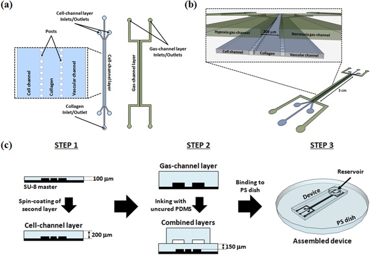
(a) Schematic of photoresist master patterns. The enlarged diagram shows the 50 × 50 μm posts along cell-channel layer length. (b) Isometric view of combined cell- and gas-channel layers. The enlarged diagram shows a cross-section indicating the cell and vascular channels and the gas channels used for delivery of the hypoxia and normoxia gas mixtures. (c) Schematic of the device fabrication process. Step 3 displays the microfluidic device bound to PS dish and the circular reservoir bound atop of the device to hold cell medium for withdrawal and maintenance of the cells during culture.

(a) Oxygen concentrations at steady-state within cross-section of the microfluidic device bound to PS dish. The oxygen content in the hypoxia and normoxia channels is 1% and 21% respectively. Scale bar marks 400 μm. (b) Predicted oxygen partial pressure gradient across the width of cell-channel layer of device bound directly to a PS dish. The theoretical oxygen gradient was obtained with a line profile at the interface between the device and the PS (represented by the dotted white line).

(a) Map of oxygen concentrations at steady-state within cross-section of microfluidic device bound to 200 μm PDMS film containing the oxygen-sensing microparticles. The oxygen content in the hypoxia and normoxia channels is 1% and 21%, respectively. Scale bar marks 400 μm. (b) Predicted oxygen partial pressure gradient across the width of cell-channel layer of device bound microparticle containing PDMS film. As with the PS model, a line profile was used at the interface between the device and the film (represented by top line of the dotted rectangle marking the microparticle film) to create this figure.

Fluorescence images of oxygen-sensing microparticles embedded within 200 μm PDMS film for (a) 0% O2 and (b) at 21% O2 at 6.4× magnification. Scale bar marks 200 μm. (c) Calibration of microparticle film using two-site Stern–Volmer model (R2 = 0.9979). Calibration data shown as mean ± standard error of the mean for the three replicates (n = 3). (d) Reversibility response of oxygen-sensing microparticles within the PDMS film. Reversibility data shown as mean ± standard deviation for the three replicates (n = 3).

(a) Fluorescence image of the spatial gradient in fluorescence observed from the oxygen-sensing microparticles embedded within the 200 μm PDMS film at 6.4× magnification. The gradient was created by infusing 1% O2 through the hypoxia channel and 21% O2 through the normoxia channel. Scale bar marks 200 μm. (b)–(d) Comparison between predicted oxygen partial pressure gradients (dashed lines) and empirical characterizations (data points) across the width of the cell-channel layer of the device bound to the PDMS film containing the oxygen-sensing microparticles for (b) 1% O2, (c) 2% O2, and (d) 5% O2. Measured gradient data shown as mean ± standard deviation for the three replicates (n = 3).

Phase contrast image of PANC-1 cells within microfluidic device during culture under (a) air/5% CO2 for 8 days and (b) 1%/21% O2 gradient for 8 days at 10× magnification. Scale bar marks 100 μm. Cells were stained with crystal violet to enhance their contrast against the transparent background of the device. The dye also stained the collagen barrier. In (b), the image shows cell migration in response to the created gradient, as well as breakage of the collagen barrier due to the cells' invasion.
Similar articles
-
A microfluidic oxygen sink to create a targeted cellular hypoxic microenvironment under ambient atmospheric conditions.Acta Biomater. 2018 Jun;73:167-179. doi: 10.1016/j.actbio.2018.04.007. Epub 2018 Apr 9. Acta Biomater. 2018. PMID: 29649636
-
Microfluidic device to attain high spatial and temporal control of oxygen.PLoS One. 2018 Dec 20;13(12):e0209574. doi: 10.1371/journal.pone.0209574. eCollection 2018. PLoS One. 2018. PMID: 30571786 Free PMC article.
-
A polydimethylsiloxane-polycarbonate hybrid microfluidic device capable of generating perpendicular chemical and oxygen gradients for cell culture studies.Lab Chip. 2014 Oct 7;14(19):3762-72. doi: 10.1039/c4lc00732h. Lab Chip. 2014. PMID: 25096368
-
HIF expression and the role of hypoxic microenvironments within primary tumours as protective sites driving cancer stem cell renewal and metastatic progression.Carcinogenesis. 2013 Aug;34(8):1699-707. doi: 10.1093/carcin/bgt209. Epub 2013 Jun 5. Carcinogenesis. 2013. PMID: 23740838 Review.
-
Cycling hypoxia: A key feature of the tumor microenvironment.Biochim Biophys Acta. 2016 Aug;1866(1):76-86. doi: 10.1016/j.bbcan.2016.06.004. Epub 2016 Jun 22. Biochim Biophys Acta. 2016. PMID: 27343712 Review.
Cited by
-
Differential Oxygenation in Tumor Microenvironment Modulates Macrophage and Cancer Cell Crosstalk: Novel Experimental Setting and Proof of Concept.Front Oncol. 2019 Feb 6;9:43. doi: 10.3389/fonc.2019.00043. eCollection 2019. Front Oncol. 2019. PMID: 30788287 Free PMC article.
-
Generating linear oxygen gradients across 3D cell cultures with block-layered oxygen controlled chips (BLOCCs).Anal Methods. 2020 Jan 7;12(1):18-24. doi: 10.1039/C9AY01690B. Epub 2019 Nov 26. Anal Methods. 2020. PMID: 32190125 Free PMC article.
-
A Relatively Small Gradient of Extracellular pH Directs Migration of MDA-MB-231 Cells In Vitro.Int J Mol Sci. 2020 Apr 7;21(7):2565. doi: 10.3390/ijms21072565. Int J Mol Sci. 2020. PMID: 32272744 Free PMC article.
-
The progressive trend of modeling and drug screening systems of breast cancer bone metastasis.J Biol Eng. 2024 Feb 5;18(1):14. doi: 10.1186/s13036-024-00408-5. J Biol Eng. 2024. PMID: 38317174 Free PMC article. Review.
-
Measuring and regulating oxygen levels in microphysiological systems: design, material, and sensor considerations.Analyst. 2019 May 13;144(10):3190-3215. doi: 10.1039/c8an02201a. Analyst. 2019. PMID: 30968094 Free PMC article. Review.
References
-
- Jessup J. M., Battle P., Waller H., Edmiston K. H., Stolz D. B., Watkins S. C., Locker J., and Skena K., Cancer Res. 59, 1825–1829 (1999). - PubMed
Grants and funding
LinkOut - more resources
Full Text Sources
Other Literature Sources