. 2015 Jan 27;9(1):860-8.
doi: 10.1021/nn506544b. Epub 2015 Jan 13.

Highly angiogenic peptide nanofibers

Affiliations

Highly angiogenic peptide nanofibers

Vivek A Kumar et al. ACS Nano. .

Abstract

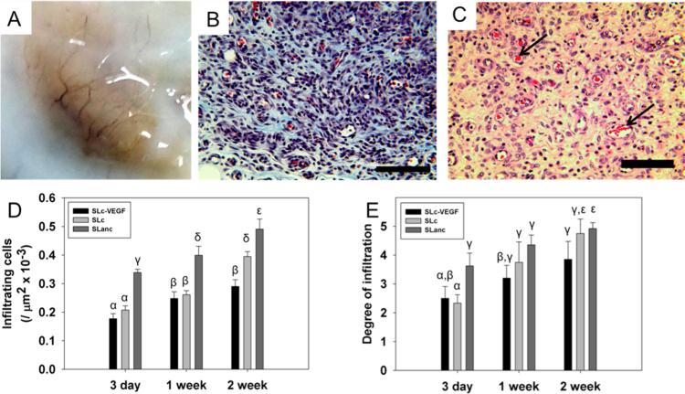
Major limitations of current tissue regeneration approaches using artificial scaffolds are fibrous encapsulation, lack of host cellular infiltration, unwanted immune responses, surface degradation preceding biointegration, and artificial degradation byproducts. Specifically, for scaffolds larger than 200-500 μm, implants must be accompanied by host angiogenesis in order to provide adequate nutrient/waste exchange in the newly forming tissue. In the current work, we design a peptide-based self-assembling nanofibrous hydrogel containing cell-mediated degradation and proangiogenic moieties that specifically address these challenges. This hydrogel can be easily delivered by syringe, is rapidly infiltrated by cells of hematopoietic and mesenchymal origin, and rapidly forms an extremely robust mature vascular network. Scaffolds show no signs of fibrous encapsulation and after 3 weeks are resorbed into the native tissue. These supramolecular assemblies may prove a vital paradigm for tissue regeneration and specifically for ischemic tissue disease.

Keywords: angiogenesis; multidomain peptide; self-assembly; supramolecular chemistry.


Figures

Figure 1
Figure 1
Physical characterization of SLanc. (A) Multidomain peptides were engineered to self-assemble, biodegrade, and present bioactive moieties. (B) SLanc peptides form a viscous solution in 298 mM sucrose (C) that gel upon the addition of anions. (D) Fluorescent carboxyfluorescein-modified SLanc (F-SLanc) was added to SLanc (1:100) and demonstrated facile gelation. Rheometry of 1 wt % gels showed high G′ and G′′ with shear thinning at high (E) strain rates and (F) high-frequency oscillation. (G) Demonstration of recovery from high shear rate, as experienced when aspirated or injected via a needle, of SLanc hydrogels. (H) FTIR spectrum shows characteristic amide I band (1625 cm −1 peak) and antiparallel (1695 cm −1 peak) β-sheet formation. (I) Circular dichroism shows the presence of a β-sheet supramolecular structure within a polymer structure (solid line), which is enhanced by the addition of polyvalent salts (dotted line). (J) SEM (scale bar 1 μm, inset 10 μm) and (K) TEM (scale bar 100 nm) show the nanofibrous matrix structure. For physical characterization of previously published MDPs including SL, SLc, and SLac, the reader is directed to refs – and –.
Figure 2
Figure 2
In vitro biochemical response. Controlled degradation of SLanc shown by MALDI mass spectrometry of intact SLanc before (A) and cleavage fragments after incubation with MMP-2 (B). Activation of VEGF receptors shown by PCR of VEGFR-1/2 and NP-1 interaction with different peptides or VEGF positive control. Similar Greek letters indicate no statistically significant difference for each receptor (*p < 0.01).
Figure 3
Figure 3
Cell adhesion and scratch wound healing on scaffolds. With respect to hMSCs, (A) TCP showed greatest adhesion, while RGD-modified SLac and angiogenic SLanc showed similar cell adhesion. Representative images of hMSC adhered and spread on all scaffolds: (B) TCP, (C) RTT, (D) SLac, (E) SLanc, and (F) SL; scale bar 100 μm. Inflammatory potential of scaffolds measured by incubating THP-1 cells atop scaffolds and measuring TNF-α (G) and IL-1β (H) secretion. Quantification of HUVEC adhesion on scaffolds showed (I) HUVEC proliferated to a similar extent on all hydrogel material surfaces after 4 days. Migration of HUVEC into a scratch wound with a soluble peptide stimulus was measured (J). Conditions were in low FBS (0.5%) media after 18 h. SLac and SLanc showed significantly higher proliferation than TCP. Representative images of a SLanc healed scratch wound are shown (K) before and (L) after; scale bar 250 μm. Similar Greek letters indicate no statistically significant difference (*p < 0.01).
Figure 4
Figure 4
Cellular infiltration and angiogenesis within scaffolds. (A) Upon explant, SLanc scaffolds showed visible macroscale vessels at 7 days. (B) Masson's Trichrome and (C) H&E staining show rapid infiltration of scaffolds and presence of blood vessels with red blood cells (arrows) at 1 week; scale bar 100 μm. A magnified image of the blood vessel clearly showing RBCs flowing through is shown in Figure S3. Control and 3 day time points shown in Figures S1 and S2. (D) SLanc scaffolds show significantly greater cellular infiltrate toward the center of scaffolds and (E) degree of infiltration, compared to SLc or SLc-VEGF. Similar Greek letters indicate no statistically significant difference (p < 0.01).
Figure 5
Figure 5
In vivo efficacy of SLanc in promoting angiogenesis, venulo/arteriolo genesis. (A) Immunostaining of cells from hematopoietic and mesenchymal origin showing extensive infiltration of pericyte-like cells (purple, Nestin+), which costain with SMC (red, α-SMA+), surrounding endothelial cells in large stable microvessels and circulating cells (green, CD31+); select region magnified in Figure S4 and controls in S5. (B) Perfusion of vessel was confirmed by observation of circulating cells of hematopoietic origin (purple, CD45+) in endothelial lined (green, vWF+) vessels. (C) Vascular tube formation and (D) vessel maturity were significantly higher in SLanc compared to SLc/SLc-VEGF controls. Similar Greek letters indicate no statistically significant difference (p < 0.01).
Figure 6
Figure 6
Proposed mechanism of healing. (A) SLanc scaffolds form nanofibrous hydrogels. (B) Scaffolds rapidly infiltrate with macrophages. (C) Hydrophobic and ionically bound SLanc at the periphery of scaffolds recruits endothelial cells to infiltrated scaffolds, which interact with current infiltrate to form blood vessels. (D) Subcutaneous and intramuscular implanted scaffolds rapidly form CD31+, vWF+, Nestin+, and α-SMA+ microvessels that have CD45+ cells that flow through their lumen. Images above (D) show progression of vessels from immature (EC only) to stabilized (EC + pericyte) to mature (EC + pericyte + SMC). (E) Neovessels in the subcutaneous model resorb since they do not anastamose injured/damaged vessels

References

    1. Radecki RP. Acute Ischemic Stroke and Timing of Treatment. JAMA. 2013;310:1855–1856. - PubMed
    1. Heusch G, Libby P, Gersh B, Yellon D, Bohm M, Lopaschuk G, Opie L. Cardiovascular Remodelling in Coronary Artery Disease and Heart Failure. Lancet. 2014;383:1933–1943. - PMC - PubMed
    1. Courties G, Moskowitz MA, Nahrendorf M. The Innate Immune System after Ischemic Injury: Lessons To Be Learned from the Heart and Brain. JAMA Neurol. 2014;71:233–236. - PMC - PubMed
    1. Sun Q, Silva EA, Wang A, Fritton JC, Mooney DJ, Schaffler MB, Grossman PM, Rajagopalan S. Sustained Release of Multiple Growth Factors from Injectable Polymeric System as a Novel Therapeutic Approach towards Angiogenesis. Pharm. Res. 2010;27:264–271. - PMC - PubMed
    1. Mohsin S, Wu JC, Sussman MA. Predicting the Future with Stem Cells. Circulation. 2014;129:136–138. - PMC - PubMed

Publication types

MeSH terms