Comparative Study
. 2015 May 15;24(10):1182-93.
doi: 10.1089/scd.2014.0486. Epub 2015 Feb 26.

Therapeutic potential of gingival fibroblasts for cutaneous radiation syndrome: comparison to bone marrow-mesenchymal stem cell grafts

Affiliations
Comparative Study

Therapeutic potential of gingival fibroblasts for cutaneous radiation syndrome: comparison to bone marrow-mesenchymal stem cell grafts

Christine Linard et al. Stem Cells Dev. .

Abstract

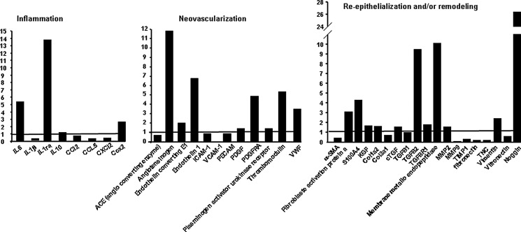
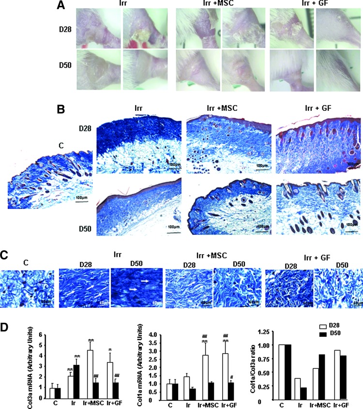
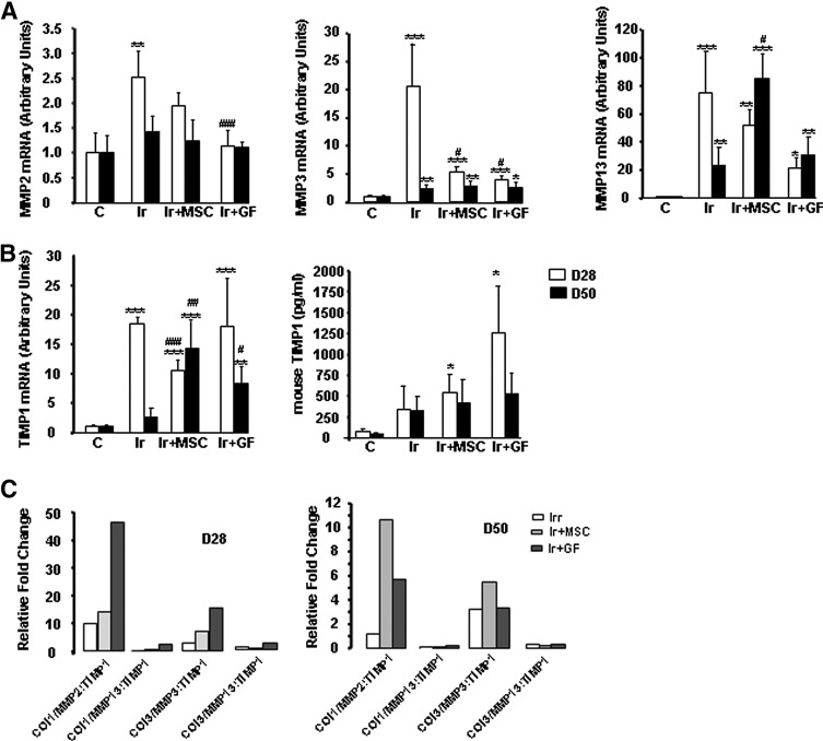
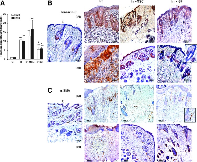
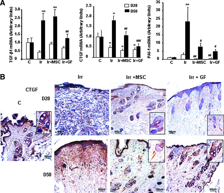
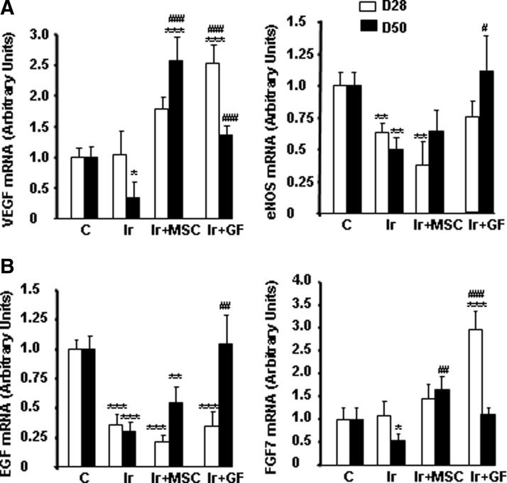
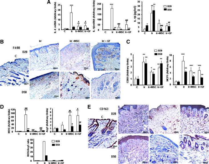
Mesenchymal stem cell (MSC) therapy has recently been investigated as a potential treatment for cutaneous radiation burns. We tested the hypothesis that injection of local gingival fibroblasts (GFs) would promote healing of radiation burn lesions and compared results with those for MSC transplantation. Human clinical- grade GFs or bone marrow-derived MSCs were intradermally injected into mice 21 days after local leg irradiation. Immunostaining and real-time PCR analysis were used to assess the effects of each treatment on extracellular matrix remodeling and inflammation in skin on days 28 and 50 postirradiation. GFs induced the early development of thick, fully regenerated epidermis, skin appendages, and hair follicles, earlier than MSCs did. The acceleration of wound healing by GFs involved rearrangement of the deposited collagen, modification of the Col/MMP/TIMP balance, and modulation of the expression and localization of tenascin-C and of the expression of growth factors (VEGF, EGF, and FGF7). As MSC treatment did, GF injection decreased the irradiation-induced inflammatory response and switched the differentiation of macrophages toward an M2-like phenotype, characterized by CD163(+) macrophage infiltration and strong expression of arginase-1. These findings indicate that GFs are an attractive target for regenerative medicine, for easier to collect, can grow in culture, and promote cutaneous wound healing in irradiation burn lesions.


Figures

<b>FIG. 1.</b>
FIG. 1.
mRNA expression levels of GF compared to BM-MSCs before transplantation. Real-time PCR assays were performed to measure mRNA levels of inflammatory, neovascularization, and remodeling factors in GF compared to BM-MSCs. The fold changes were generated by comparing cultured hGF (histogramme) with hMSCs (basal line to 1). GAPDH was quantified as an internal control. MSC, mesenchymal stem cell; GF, gingival fibroblast; Irr, irradiation.
<b>FIG. 2.</b>
FIG. 2.
MSCs and GFs modified radiation-induced collagen deposition. (A) Representative macroscopic pictures, (B) Masson trichrome staining in irradiated groups, MSC-treated groups, and GF-treated groups on days 28 and 50 after irradiation. ×20 magnification. (C) Staining revealed a high density of dermal collagen fibers with unidirectional alignment parallel to the white arrows after irradiation and the restoration of multidirectional alignment after MSC or GF treatment. (D) Real-time PCR of Col3a, Col1a, and Col1a/Col3a ratio. Data are expressed relative to control mice and normalized to GAPDH. Results are expressed as mean±SEM. P values were calculated by analysis of variance (ANOVA) with Bonferroni correction, *P<0.05; **P<0.01 compared with nonirradiated controls; #P<0.05; ##P<0.01 compared with irradiated-untreated controls.
<b>FIG. 3.</b>
FIG. 3.
MSC or GF treatment influences ECM remodeling. (A) Real-time expression of MMP-2, MMP-3, and MMP-13. (B) TIMP-1 mRNA and protein expression. Levels of mRNA are expressed relative to control mice and normalized to GAPDH. Protein was measured in skin by ELISA test. (C) Collagen-to-MMP-to-TIMP ratio: relative expression of MMP was calculated by determining the fold change of MMP mRNA levels relative to its relevant inhibitor TIMP in irradiated-untreated and irradiated MSC- and GF-treated groups on days 28 and 50 after irradiation, compared with control groups. Results are expressed as mean±SEM. P values were calculated by ANOVA with Bonferroni correction, *P<0.05; **P<0.01; ***P<0.001 compared with nonirradiated controls; #P<0.05; ##P<0.01; ###P<0.001 compared with irradiated-untreated controls. ECM, extracellular matrix.
<b>FIG. 4.</b>
FIG. 4.
Expression of ECM components. (A) Real-time PCR of tenascin-C in nonirradiated control skin, irradiated-untreated skin, and MSC- or GF-treated skin on days 28 and 50 after irradiation. Data are reported relative to control mice and normalized to GAPDH. (B) Representative immunohistochemical staining with the primary anti-tenascin-C antibody and (C) the primary α-SMA antibody. Inserts were amplified local areas from the same images. Original magnification ×20. Results are expressed as mean±SEM. P values were calculated by ANOVA with Bonferroni correction, **P<0.001; compared with nonirradiated controls; #P<0.01 compared with irradiated-untreated controls.
<b>FIG. 5.</b>
FIG. 5.
Expression of wound healing-related factors. (A) Real-time PCR of TGF-β, CTGF, and PAI-1 in nonirradiated control skin, in irradiated-untreated skin, and in MSC- or GF-treated skin on days 28 and 50 after irradiation. Data are reported relative to control mice and normalized to GAPDH. (B) Representative immunohistochemical staining with the primary CTGF antibody. Inserts were amplified local areas from the same images. Original magnification ×20. Results are expressed as mean±SEM. P values were calculated by ANOVA with Bonferroni correction, *P<0.05; **P<0.01 compared with nonirradiated controls; #P<0.05; ##P<0.01; ###P<0.001 compared with irradiated-untreated controls.
<b>FIG. 6.</b>
FIG. 6.
Expression of growth factors implicated in wound repair. Real-time PCR of VEGF and eNOS (A) and EGF and FGF7 (B) in nonirradiated control skin, in irradiated-untreated skin, and in MSC- or GF-treated skin on days 28 and 50 after irradiation. Data are reported relative to control mice and normalized to GAPDH. Results are expressed as mean±SEM. P values were calculated by ANOVA with Bonferroni correction, *P<0.05; **P<0.01; ***P<0.001 compared with nonirradiated controls; #P<0.05; ##P<0.01; ###P<0.001 compared with irradiated-untreated controls. eNOS, endothelial nitric oxide synthase.
<b>FIG. 7.</b>
FIG. 7.
Modification of inflammatory profile by MSC or GF treatment. (A) Real-time-PCR analysis of IL-6 and IL-1β. Protein level of IL-1β was analyzed in skin by ELISA test. (B) Representative immunostaining for F4/80-positive macrophages. (C) Real-time PCR analysis of CD68, MCP1, genes related to macrophage infiltration. (D) Real-time PCR analysis of genes related to M1 macrophage iNOS and M2 macrophage activity Arg-1 and iNOS/Arg-1 mRNA level ratio as an index of M1/M2 activity balance. (E) Representative immunostaining of CD163. Original magnification ×20. Results are expressed as mean±SEM. P values were calculated by ANOVA with Bonferroni correction, *P<0.05; **P<0.01; ***P<0.001 compared with nonirradiated controls; #P<0.05; ##P<0.01; ###P<0.001 compared with irradiated untreated controls. iNOS, inducible nitric oxide synthase.

References

    1. Williams JP. and McBride WH. (2011). After the bomb drops: a new look at radiation-induced multiple organ dysfunction syndrome (MODS). Int J Radiat Biol 87:851–868 - PMC - PubMed
    1. Peter RU. (2005). Cutaneous radiation syndrome in multi-organ failure. BJR (Suppl. 27):180–184
    1. Hill RP, Rodemann HP, Hendry JH, Roberts SA. and Anscher MS. (2001). Normal tissue radiobiology: from the laboratory to the clinic. Int J Radiat Oncol Biol Phys 49:353–365 - PubMed
    1. Wu Y, Wang J, Scott PG. and Tredget EE. (2007). Bone marrow-derived stem cells in wound healing: a review. Wound Repair Regen 15 (Suppl. 1):S18–S26 - PubMed
    1. Lataillade JJ, Doucet C, Bey E, Carsin H, Huet C, Clairand I, Bottollier-Depois JF, Chapel A, Ernou I, et al. (2007). New approach to radiation burn treatment by dosimetry-guided surgery combined with autologous mesenchymal stem cell therapy. Regen Med 2:785–794 - PubMed

Publication types