. 2015 Jun 5;427(11):2056-71.
doi: 10.1016/j.jmb.2015.01.001. Epub 2015 Jan 10.

Proteomic characterization of the nucleolar linker histone H1 interaction network

Affiliations

Proteomic characterization of the nucleolar linker histone H1 interaction network

Heather J Szerlong et al. J Mol Biol. .

Abstract

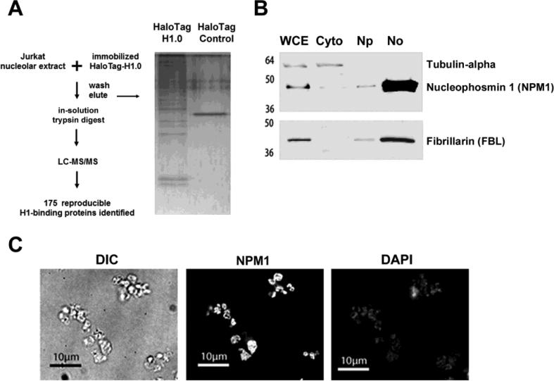
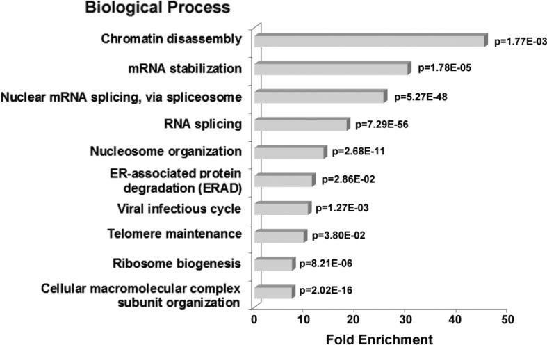
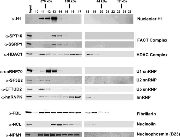
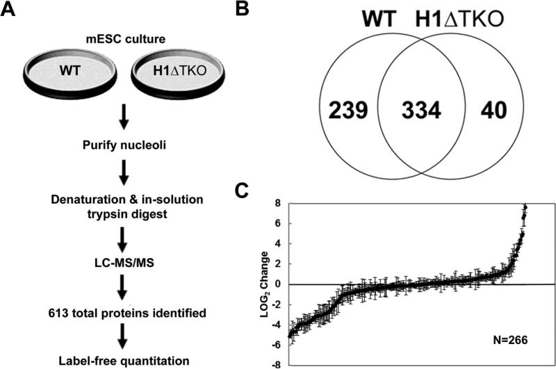
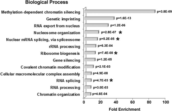
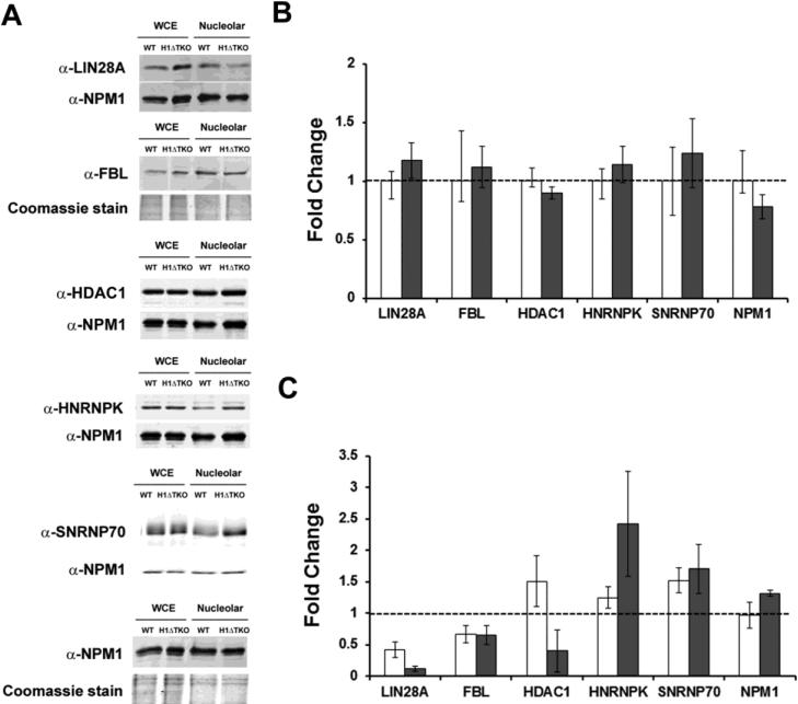
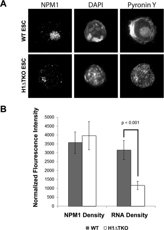
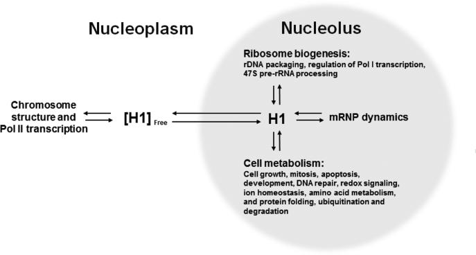
To investigate the relationship between linker histone H1 and protein-protein interactions in the nucleolus, we used biochemical and proteomics approaches to characterize nucleoli purified from cultured human and mouse cells. Mass spectrometry identified 175 proteins in human T cell nucleolar extracts that bound to Sepharose-immobilized H1 in vitro. Gene ontology analysis found significant enrichment for H1 binding proteins with functions related to nucleolar chromatin structure and RNA polymerase I transcription regulation, rRNA processing, and mRNA splicing. Consistent with the affinity binding results, H1 existed in large (400 to >650kDa) macromolecular complexes in human T cell nucleolar extracts. To complement the biochemical experiments, we investigated the effects of in vivo H1 depletion on protein content and structural integrity of the nucleolus using the H1 triple isoform knockout (H1ΔTKO) mouse embryonic stem cell (mESC) model system. Proteomic profiling of purified wild-type mESC nucleoli identified a total of 613 proteins, only ~60% of which were detected in the H1 mutant nucleoli. Within the affected group, spectral counting analysis quantitated 135 specific nucleolar proteins whose levels were significantly altered in H1ΔTKO mESC. Importantly, the functions of the affected proteins in mESC closely overlapped with those of the human T cell nucleolar H1 binding proteins. Immunofluorescence microscopy of intact H1ΔTKO mESC demonstrated both a loss of nucleolar RNA content and altered nucleolar morphology resulting from in vivo H1 depletion. We conclude that H1 organizes and maintains an extensive protein-protein interaction network in the nucleolus required for nucleolar structure and integrity.

Keywords: chromatin structure and gene expression; mass spectrometry; messenger RNA splicing; protein–protein interactions; ribosome biogenesis.


Figures

Fig. 1
Fig. 1
Experimental design and isolation of nucleoli from cultured T cells. (a) Schematic representation of H1-affinity binding experiments and proteomics workflow (left). Purified nucleoli from Jurkat T-cells were used as the protein source for H1-binding experiments. Proteins that bound HaloTag-H1 or Halo Tag control beads were eluted, digested with trypsin protease, and identified by LC-MS/MS analysis. A portion of the eluate was resolved by SDS-PAGE and stained with silver (right). (b) Immunoblots of Jurkat T-cell subcellular fractions (5 μg protein per lane) probed with the indicated antibodies (right). The fractions (top) and molecular weight standards (left) are indicated. Whole cell extract (WCE), cytoplasm (Cyto), nucleoplasm (Np) and nucleolar (No). (c) Nucleolar fractions from Jurkat T-cells were fixed in 4% paraformaldehyde and analyzed by differential interference contrast (DIC) microscopy or indirect immunofluorescence with either α-nucleophosmin antibody or stained with 4',6-Diamidino-2-Phenylindole, Dihydrochloride (DAPI).
Fig. 2
Fig. 2
GO biological process enrichment in nucleolar H1 binding proteins identified by LC-MS/MS analysis. The bar graph represents the fold enrichment of GO biological processes terms in H1 binding proteins relative to the Human gene database using the DAVID Bioinformatics Resources functional annotation clustering algorithm (31). The most highly enriched functions were chosen from a cluster of similar terms. Biological processes with <4-fold enrichment are not shown. A p-value for each term is indicated.
Fig. 3
Fig. 3
Size exclusion chromatography (SEC) analysis of purified nucleoli. Immunoblot analysis of nucleolar extract from Jurkat T-cells resolved by SEC under native buffer conditions. Purified nucleoli were sonicated and treated with DNAse I in high salt (0.5 M NaCl) prior to loading on a S200 Sepharose column pre-equilibrated in low salt (200 mM NaCl). Individual fractions (top) were resolved by SDS-PAGE, transferred to PVDF membrane and probed with the indicated primary antibodies (left). The apparent MW range for each protein or protein complex (right) was extrapolated from a standard curve derived from MW standards (Table I).
Fig. 4
Fig. 4
Comparative proteomic profile of nucleoli from WT and H1ΔTKO mESC. (a) Schematic representation of sample preparation from either WT or H1ΔTKO mESC and proteomics workflow. Purified nucleoli were treated with nuclease, denatured and digested with trypsin for LC-MS/MS analysis. Protein identification results for biological replicates were compiled for quantitative spectral counting analysis. (b) Venn diagram showing the overlap of proteins identified in WT and H1ΔTKO mESC. (c) Scatter plot representing the normalized LOG2 values of H1ΔTKO over WT control. Three biological repeats were performed for each strain and proteins identified in at least two out of the three samples with a sum of >10 spectra and standard deviations below the average standard deviation of the experiment were included in the analysis. Negative LOG2 values indicate reduced protein levels whereas positive LOG2 values represent elevated protein levels in H1ΔTKO relative to WT mESC.
Fig. 5
Fig. 5
GO Biological process enrichment in proteins altered in H1ΔTKO mESC. The bar graph shows the fold enrichment of GO biological processes terms in the H1-depleted dataset relative to the Mus musculus database using the DAVID Bioinformatics Resources functional annotation clustering algorithm (31). The most highly enriched functions were chosen from a cluster of similar terms. A p-value for each term is indicated. Asterisks (*) indicated terms shared with the H1-binding proteins identified in T-cells (Fig. 2.).
Fig. 6
Fig. 6
Expression of H1ΔTKO mESC proteins and validation of SpC analysis. (a) Immunoblot analysis of extracts from either WT or H1ΔTKO mESC was used to determine the relative abundance of proteins identified by SpC analysis in either the whole cell (b) or in the nucleolus (c). Five μg of total protein from each sample was resolved by SDS-PAGE and probed for the indicated proteins (left). As a loading control, the same immunoblot was either probed with α-NPM1 or the gel was stained with coomassie. (b) The fold-change expression in mESC whole cell extracts was determined by dividing the normalized signal for H1ΔTKO (closed bars) by WT (open bars) set at 1.0 for each of the indicated proteins. (c) The fold-change expression of candidate proteins in nucleolar extracts was determined by dividing the normalized values for H1ΔTKO by WT for each of the indicted proteins. Bar graphs represent the values obtained from either immunoblot analysis (open bars) or SpC analysis (closed bars). Values are the averages ± the standard deviations of triplicate samples and normalized to the loading control present in each lane.
Fig. 7
Fig. 7
Immunofluorescence microscopy of WT and H1ΔTKO mESC. (a) High-resolution immunofluorescence images of mESC stained with NPM1 antibody, DAPI and Pyronin Y. B, The fluorescence intensity for NPM1 and Pyronin Y was plotted for WT (closed bar) and H1ΔTKO (closed bar) mESC. Fluorescence signal was normalized to nucleolar area in each cell. Error bars represent the standard deviation among >25 cells. Pyronin Y (RNA) signal density deceases in H1ΔTKO mESC (student t-test p<0.001).
Fig. 8
Fig. 8
Model for a nucleolar H1 protein-protein interaction network (PPIN). H1 transitions between the nucleoplasm and the nucleolus to regulate the dynamics of proteins associated with the H1 protein-protein interaction network (PPIN). For nuclear genes, a major role for H1 is to modulate Pol II transcription. In the nucleolus, the H1 PPIN functions in various aspects of ribosome biogenesis, pre-mRNA splicing (or mRNP dynamics), and an assortment of other functions related to cellular health and disease. Through these interactions, H1 functions as a hub protein that directly impacts the nuclear steady-state equilibrium and accumulation of proteins and RNP complexes in the nucleolus.

References

    1. Happel N, Doenecke D. Histone H1 and its isoforms: contribution to chromatin structure and function. Gene. 2009;431:1–12. - PubMed
    1. Kowalski A, Palyga J. Linker histone subtypes and their allelic variants. Cell biology international. 2012;36:981–996. - PubMed
    1. Izzo A, Kamieniarz K, Schneider R. The histone H1 family: specific members, specific functions? Biological chemistry. 2008;389:333–343. - PubMed
    1. Catez F, Ueda T, Bustin M. Determinants of histone H1 mobility and chromatin binding in living cells. Nat Struct Mol Biol. 2006;13:305–310. - PMC - PubMed
    1. Luger K, Mader AW, Richmond RK, Sargent DF, Richmond TJ. Crystal structure of the nucleosome core particle at 2.8 A resolution. Nature. 1997;389:251–260. - PubMed

Publication types

LinkOut - more resources