

Multi-frequency intravascular ultrasound (IVUS) imaging
- PMID: 25585394
- PMCID: PMC4522164
- DOI: 10.1109/TUFFC.2014.006679
Multi-frequency intravascular ultrasound (IVUS) imaging
Erratum in
-
Erratum: Multi-frequency intravascular ultrasound (IVUS) imaging.IEEE Trans Ultrason Ferroelectr Freq Control. 2015 Mar;62(3):604. doi: 10.1109/TUFFC.2015.007057. IEEE Trans Ultrason Ferroelectr Freq Control. 2015. PMID: 25768827 No abstract available.
Abstract
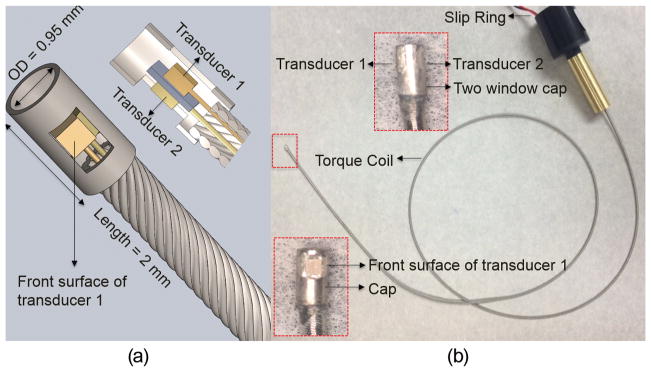
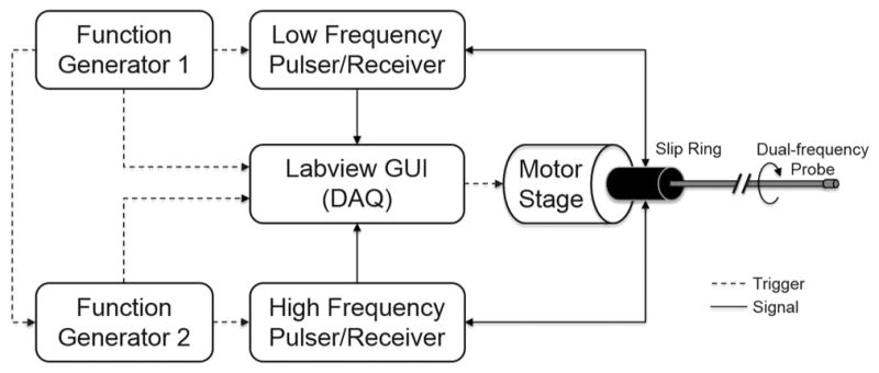
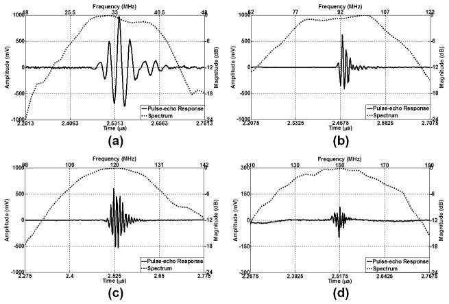
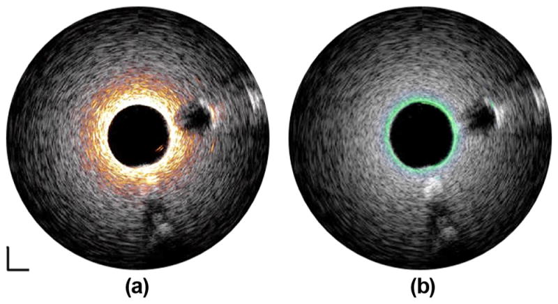
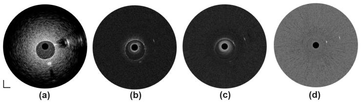
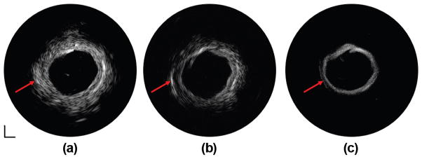
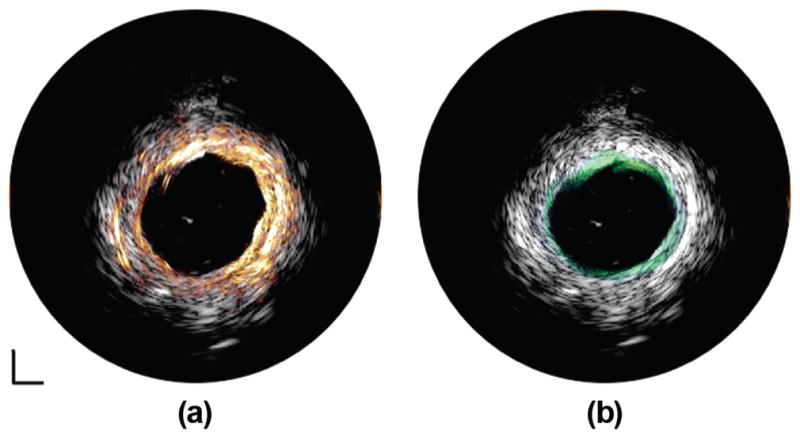
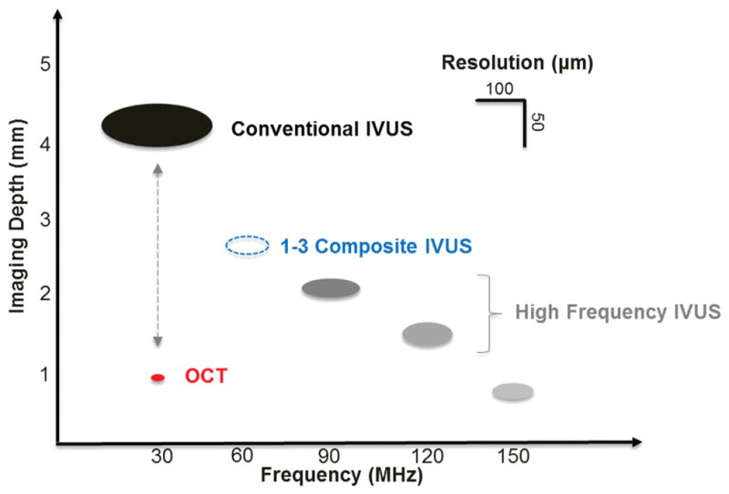
Acute coronary syndrome (ACS) is frequently associated with the sudden rupture of a vulnerable atherosclerotic plaque within the coronary artery. Several unique physiological features, including a thin fibrous cap accompanied by a necrotic lipid core, are the targeted indicators for identifying the vulnerable plaques. Intravascular ultrasound (IVUS), a catheter-based imaging technology, has been routinely performed in clinics for more than 20 years to describe the morphology of the coronary artery and guide percutaneous coronary interventions. However, conventional IVUS cannot facilitate the risk assessment of ACS because of its intrinsic limitations, such as insufficient resolution. Renovation of the IVUS technology is essentially needed to overcome the limitations and enhance the coronary artery characterization. In this paper, a multi-frequency intravascular ultrasound (IVUS) imaging system was developed by incorporating a higher frequency IVUS transducer (80 to 150 MHz) with the conventional IVUS (30-50 MHz) system. The newly developed system maintains the advantage of deeply penetrating imaging with the conventional IVUS, while offering an improved higher resolution image with IVUS at a higher frequency. The prototyped multifrequency catheter has a clinically compatible size of 0.95 mm and a favorable capability of automated image co-registration. In vitro human coronary artery imaging has demonstrated the feasibility and superiority of the multi-frequency IVUS imaging system to deliver a more comprehensive visualization of the coronary artery. This ultrasonic-only intravascular imaging technique, based on a moderate refinement of the conventional IVUS system, is not only cost-effective from the perspective of manufacturing and clinical practice, but also holds the promise of future translation into clinical benefits.
Figures

References
-
- Virmani R, Kolodgie FD, Burke AP, Farb A, Schwartz SM. Lessons from sudden coronary death: A comprehensive morphological classification scheme for atherosclerotic lesions. Arterioscler Thromb Vasc Biol. 2000 May;20:1262–1275. - PubMed
-
- Kolodgie FD, Burke AP, Farb A, Gold HK, Yuan J, Narula J, Finn AV, Virmani R. The thin-cap fibroatheroma: A type of vulnerable plaque: The major precursor lesion to acute coronary syndromes. Curr Opin Cardiol. 2001 Sep;16:285–292. - PubMed
-
- Garcia-Garcia HM, Gonzalo N, Granada JF, Regar E, Serruys PW. Diagnosis and treatment of coronary vulnerable plaques. Expert Rev Cardiovasc Ther. 2008 Feb;6:209–222. - PubMed
-
- Puri R, Worthley MI, Nicholls SJ. Intravascular imaging of vulnerable coronary plaque: Current and future concepts. Nat Rev Cardiol. 2011 Mar;8:131–139. - PubMed
-
- Potkin BN, Bartorelli AL, Gessert JM, Neville RF, Almagor Y, Roberts WC, Leon MB. Coronary artery imaging with intravascular high-frequency ultrasound. Circulation. 1990 May;81:1575–1585. - PubMed
Publication types
MeSH terms
Grants and funding
LinkOut - more resources
Full Text Sources
Other Literature Sources
Miscellaneous
