

S100P, a calcium-binding protein, is preferentially associated with the growth of polypoid tumors in colorectal cancer
- PMID: 25585623
- PMCID: PMC4314409
- DOI: 10.3892/ijmm.2015.2065
S100P, a calcium-binding protein, is preferentially associated with the growth of polypoid tumors in colorectal cancer
Abstract
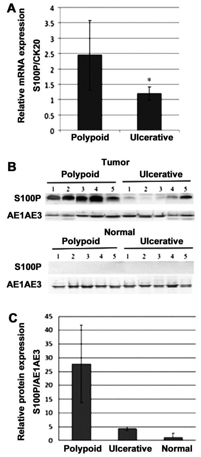
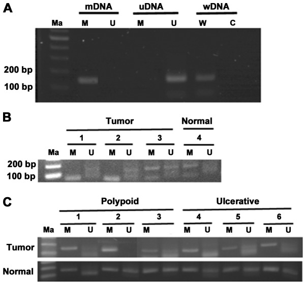
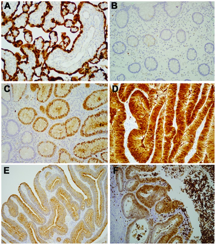
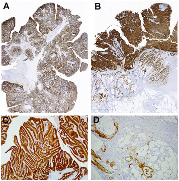
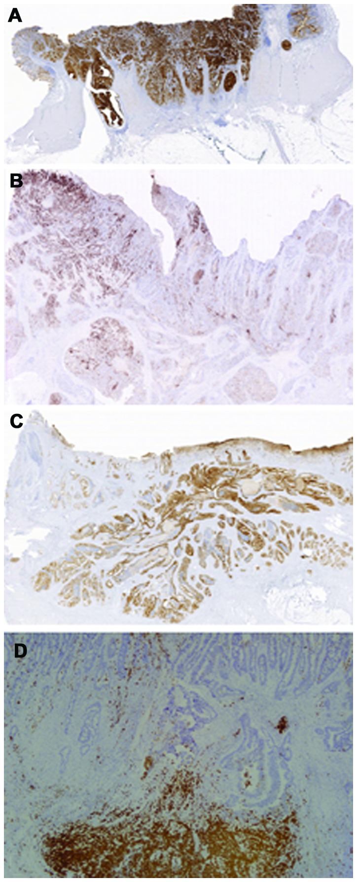
Colorectal cancer (CRC) is a genetically heterogeneous disease with distinct morphological patterns. It has been shown that polypoid and ulcerative CRC displays different genetic alterations. In the present study, we aimed to investigate genes with differential expression patterns between ulcerative and polypoid CRC. cDNA microarray analysis was performed to compare the gene expression profiles in samples of ulcerative and polypoid CRC with paired normal mucosa samples. Potential candidate genes were further validated using reverse transcription-quantitative polymerase chain reaction (RT-qPCR), western blot analysis and immunohistochemistry. The epigenetic regulation of gene expression was investigated using methylation-specific PCR (MSP). cDNA microarray analysis identified 11 upregulated and 14 downregulated genes which were differentially expressed in samples from both tumor types compared to the matched normal mucosa samples. Among these, S100P was the only upregulated gene preferentially associated with polypoid CRC (P=0.032). The samples of polypoid CRC displayed significantly higher S100P protein and mRNA expression levels than the samples of ulcerative CRC (P<0.05, respectively). Using semi-quantitative immunohistochemical analyses, S100P overexpression was found to be preferentially associated with polypoid CRC (24/30 vs. 14/40, P<0.001). The relative methylation level determined by MSP did not differ significantly between the samples of polypoid and ulcerative CRC (43.36 vs. 49.10%, P=0.168), indicating that promoter hypomethylation was not directly related to the upregulation of S100P mRNA. Our results demonstrate that the upregulation of S100P mRNA and protein expression is a predominant characteristic in polypoid CRC, whereas ulcerative CRC presents with a wide range of expression levels, indicating that S100P overexpression is not a key determinant in conferring invasion properties. The clinicopathological significance of S100P in CRC requires further investigation in well-controlled studies.
Figures
Similar articles
-
S100P, a potential novel prognostic marker in colorectal cancer.Oncol Rep. 2012 Jul;28(1):303-10. doi: 10.3892/or.2012.1794. Epub 2012 Apr 30. Oncol Rep. 2012. PMID: 22552710
-
Thioredoxin-1 promotes colorectal cancer invasion and metastasis through crosstalk with S100P.Cancer Lett. 2017 Aug 10;401:1-10. doi: 10.1016/j.canlet.2017.04.036. Epub 2017 May 5. Cancer Lett. 2017. PMID: 28483515
-
Anti-Cancer Effect of α-Solanine by Down-Regulating S100P Expression in Colorectal Cancer Cells.Recent Pat Anticancer Drug Discov. 2018;13(2):240-247. doi: 10.2174/1574892813666180329163945. Recent Pat Anticancer Drug Discov. 2018. PMID: 29600769
-
S100P: a novel therapeutic target for cancer.Amino Acids. 2011 Oct;41(4):893-9. doi: 10.1007/s00726-010-0496-4. Epub 2010 May 28. Amino Acids. 2011. PMID: 20509035 Free PMC article. Review.
-
S100P, a peculiar member of S100 family of calcium-binding proteins implicated in cancer.Acta Virol. 2013;57(2):238-46. doi: 10.4149/av_2013_02_238. Acta Virol. 2013. PMID: 23600880 Review.
Cited by
-
Inhibition of lung cancer by vitamin D depends on downregulation of histidine-rich calcium-binding protein.J Adv Res. 2020 Aug 27;29:13-22. doi: 10.1016/j.jare.2020.08.013. eCollection 2021 Mar. J Adv Res. 2020. PMID: 33842001 Free PMC article.
-
S100P interacts with integrin α7 and increases cancer cell migration and invasion in lung cancer.Oncotarget. 2015 Oct 6;6(30):29585-98. doi: 10.18632/oncotarget.4987. Oncotarget. 2015. PMID: 26320193 Free PMC article.
-
Prognostic and predictive value of the macroscopic growth pattern in patients undergoing curative resection of colorectal cancer: a single-institution retrospective cohort study of 4,080 Chinese patients.Cancer Manag Res. 2018 Jul 4;10:1875-1887. doi: 10.2147/CMAR.S165279. eCollection 2018. Cancer Manag Res. 2018. PMID: 30013394 Free PMC article.
-
Diverse LEF/TCF Expression in Human Colorectal Cancer Correlates with Altered Wnt-Regulated Transcriptome in a Meta-Analysis of Patient Biopsies.Genes (Basel). 2020 May 11;11(5):538. doi: 10.3390/genes11050538. Genes (Basel). 2020. PMID: 32403323 Free PMC article.
-
Tissue mRNA for S100A4, S100A6, S100A8, S100A9, S100A11 and S100P Proteins in Colorectal Neoplasia: A Pilot Study.Molecules. 2021 Jan 14;26(2):402. doi: 10.3390/molecules26020402. Molecules. 2021. PMID: 33466593 Free PMC article.
References
Publication types
MeSH terms
Substances
LinkOut - more resources
Full Text Sources
Other Literature Sources
Medical
Molecular Biology Databases
Research Materials
