Review
. 2015 Apr;54(2):R55-73.
doi: 10.1530/JME-14-0237. Epub 2015 Jan 13.

Genetic regulation of murine pituitary development

Affiliations
Review

Genetic regulation of murine pituitary development

Karine Rizzoti. J Mol Endocrinol. 2015 Apr.

Abstract

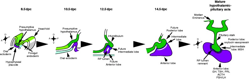
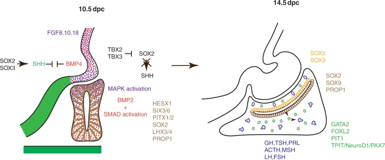
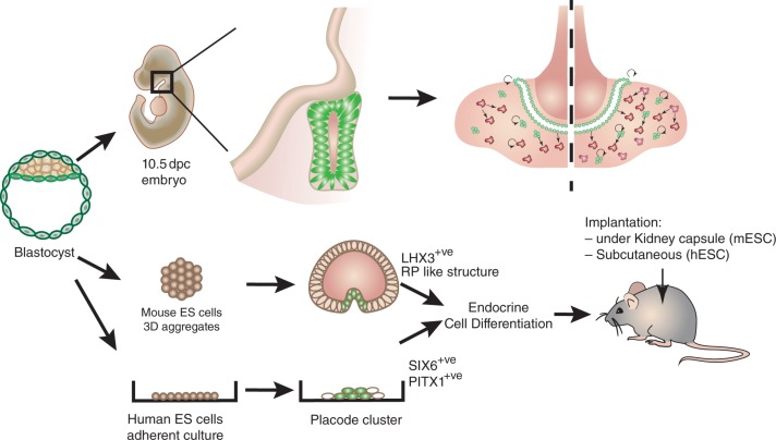
Significant progress has been made recently in unravelling the embryonic events leading to pituitary morphogenesis, both in vivo and in vitro. This includes dissection of the molecular mechanisms controlling patterning of the ventral diencephalon that regulate formation of the pituitary anlagen or Rathke's pouch. There is also a better characterisation of processes that underlie maintenance of pituitary progenitors, specification of endocrine lineages and the three-dimensional organisation of newly differentiated endocrine cells. Furthermore, a population of adult pituitary stem cells (SCs), originating from embryonic progenitors, have been described and shown to have not only regenerative potential, but also the capacity to induce tumour formation. Finally, the successful recapitulation in vitro of embryonic events leading to generation of endocrine cells from embryonic SCs, and their subsequent transplantation, represents exciting advances towards the use of regenerative medicine to treat endocrine deficits. In this review, an up-to-date description of pituitary morphogenesis will be provided and discussed with particular reference to pituitary SC studies.

Keywords: developmental factors; morphogenesis; pituitary; stem cell.


Figures

Figure 1
Figure 1
Development and function of the pituitary–hypothalamic axis. Pituitary development is initiated with the appearance of the hypophyseal placode, apposed to the future ventral diencephalon. The placode later invaginates to become Rathke's pouch (RP), still in contact in its dorsal most part with the infundibulum, which lies within the ventral diencephalon. The infundibulum then evaginates towards RP. It will give rise to the median eminence, the pituitary stalk and the posterior lobe, while the anterior and intermediate lobes originate from RP. Postnatally, the hypothalamus centralises peripheral information and controls pituitary endocrine secretions through release of hypophysiotrophic hormones. Hypothalamic peptide hormones can reach the gland directly, such as oxytocin and vasopressin secreted directly in the posterior lobe, or via the hypophyseal portal system; peptides (GnRH, gonadotrophin-releasing hormone; GHRH, GH-releasing hormone; TRH, thyrotropin-releasing hormone; CRH, corticotropin-releasing hormone; and the inhibitory SST, somatostatin) are secreted at the median eminence and collected by capillaries (adapted from Rizzoti & Lovell-Badge (2011)). Anterior pituitary endocrine cells comprise lactotrophs (producing prolactin, Prl), gonadotrophs (producing luteinizing hormone, LH; and follicle stimulating hormone, FSH), thyrotrophs (producing thyroid stimulating hormone, TSH), corticotrophs (producing adrenocorticotrophic hormone, ACTH; proteolytically cleaved from proopiomelanocortin, POMC) and somatotrophs (producing growth hormone, GH).
Figure 2
Figure 2
Main signalling pathways and factors required during pituitary development. Opposing activities of BMP4 and SHH within the ventral diencephalon pattern this region and participate in correct morphogenesis of the infundibulum and in consequence positioning of RP. Within the infundibulum, BMP4 and FGF8, FGF10 and FGF18 are required for development of RP. In RP at 10.5 dpc, BMP2 is present and there is a uniform SMAD activation profile, while the FGF pathway is only activated dorsally (Davis et al. 2011). At this stage, different transcription factors required for RP progenitor proliferation and/or maintenance are ubiquitously expressed. Later, at 14.5 dpc, progenitors are confined around the RP lumen, while differentiating and differentiated cells away from the lumen define the developing anterior pituitary.
Figure 3
Figure 3
In vivo and in vitro regenerative potential of stem cells in the pituitary. In vivo, pituitary SCs can be stimulated by physiological challenge, proliferate and differentiate into the appropriate endocrine cell type. In vitro, different strategies have been successfully devised to obtain transplantable endocrine cells. Using mouse ES cells, Suga et al. (2011) were able to recapitulate early pituitary development and mimic RP morphogenesis in 3D aggregates (Suga et al. 2011). Endocrine cells were further differentiated. Transplantation in hypophysectomised mice demonstrated that these cells were functional. Dincer et al. (2013) were also able to differentiate endocrine cells from human ES cells, in adherent cultures, initially by inducing placodal fate, then hypophyseal identity. Transplantation demonstrated in vivo secretion. These are significant steps towards regenerative medicine to cure long-term pituitary endocrine deficiencies.

References

    1. Ai D, Wang J, Amen M, Lu MF, Amendt BA, Martin JF. Nuclear factor 1 and T-cell factor/LEF recognition elements regulate Pitx2 transcription in pituitary development. Molecular and Cellular Biology. 2007;27:5765–5775. doi: 10.1128/MCB.01848-06. - DOI - PMC - PubMed
    1. elAmraoui A, Dubois PM. Experimental evidence for the early commitment of the presumptive adenohypophysis. Neuroendocrinology. 1993;58:609–615. doi: 10.1159/000126599. - DOI - PubMed
    1. Andoniadou CL, Signore M, Sajedi E, Gaston-Massuet C, Kelberman D, Burns AJ, Itasaki N, Dattani M, Martinez-Barbera JP. Lack of the murine homeobox gene Hesx1 leads to a posterior transformation of the anterior forebrain. Development. 2007;134:1499–1508. doi: 10.1242/dev.02829. - DOI - PMC - PubMed
    1. Andoniadou CL, Signore M, Young RM, Gaston-Massuet C, Wilson SW, Fuchs E, Martinez-Barbera JP. HESX1- and TCF3-mediated repression of Wnt/β-catenin targets is required for normal development of the anterior forebrain. Development. 2011;138:4931–4942. doi: 10.1242/dev.066597. - DOI - PMC - PubMed
    1. Andoniadou CL, Matsushima D, Mousavy Gharavy SN, Signore M, Mackintosh AI, Schaeffer M, Gaston-Massuet C, Mollard P, Jacques TS, Le Tissier P, et al. Sox2(+) stem/progenitor cells in the adult mouse pituitary support organ homeostasis and have tumor-inducing potential. Cell Stem Cell. 2013;13:433–445. doi: 10.1016/j.stem.2013.07.004. - DOI - PubMed

LinkOut - more resources