. 2015 Jul;26(7):1588-96.
doi: 10.1681/ASN.2014050463. Epub 2015 Jan 13.

MicroRNA-687 Induced by Hypoxia-Inducible Factor-1 Targets Phosphatase and Tensin Homolog in Renal Ischemia-Reperfusion Injury

Affiliations

MicroRNA-687 Induced by Hypoxia-Inducible Factor-1 Targets Phosphatase and Tensin Homolog in Renal Ischemia-Reperfusion Injury

Kirti Bhatt et al. J Am Soc Nephrol. 2015 Jul.

Abstract

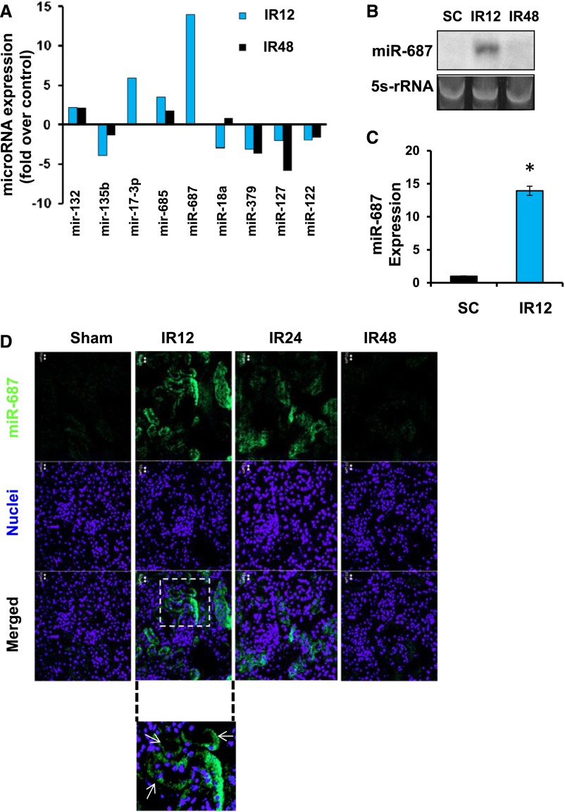
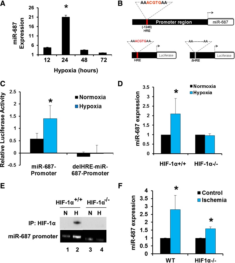
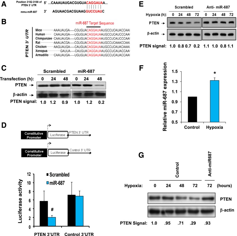
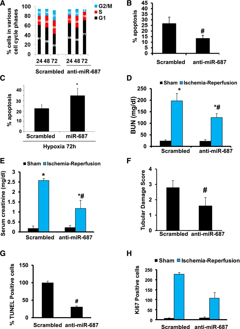
Ischemia-reperfusion injury contributes to tissue damage and organ failure in clinical settings, but the underlying mechanism remains elusive and effective therapies are still lacking. Here, we identified microRNA 687 (miR-687) as a key regulator and therapeutic target in renal ischemia-reperfusion injury. We show that miR-687 is markedly upregulated in the kidney during renal ischemia-reperfusion in mice and in cultured kidney cells during hypoxia. MiR-687 induction under these conditions was mediated by hypoxia-inducible factor-1 (HIF-1). Upon induction in vitro, miR-687 repressed the expression of phosphatase and tensin homolog (PTEN) and facilitated cell cycle progression and apoptosis. Blockade of miR-687 preserved PTEN expression and attenuated cell cycle activation and renal apoptosis, resulting in protection against kidney injury in mice. Collectively, these results unveil a novel HIF-1/miR-687/PTEN signaling pathway in ischemia-reperfusion injury that may be targeted for therapy.

Keywords: acute renal failure; cell death; hypoxia; ischemia-reperfusion; renal injury.


Figures

Figure 1.
Figure 1.
Upregulation of miR-687 during renal ischemia-reperfusion injury. (A) Significant changes in miRNA expression during renal IRI. C57BL/6 mice were subjected to 30 minutes of bilateral renal ischemia followed by 12 or 48 hours of reperfusion (IR12, IR48), while control mice had sham surgery (SC). Total RNA was extracted from kidney cortical tissues for miRNA microarray analysis. miRNAs showing consistent, significant (>2-fold over control) changes in expression during renal IRI were shortlisted. The values presented are the averages of fold changes in two separate experiments and microarray analyses. (B) Northern blot analysis of miR-687. Ten micrograms of the total RNA extracted from kidney cortex of sham-operated control mice or mice with ischemia followed by 12 or 48 hours of reperfusion were used for Northern blot analysis. 5s r-RNA was probed as loading control. (C) Real-time PCR of miR-687. RNA from kidney cortical tissues of sham-operated control or mice with renal ischemia followed by 12 or 48 hours of reperfusion were analyzed by real-time PCR to quantify miR-687. Data are the mean±SD (n=3). *P<0.05 versus SC. (D) Localization of miR-687 expression in kidney tissues 400X. Kidney tissues of sham-operated control mice or renal IRI mice were analyzed by in situ hybridization using miR-687–specific probes (green). The tissues were also exposed to DAPI to reveal nuclei (purple). Arrows: renal tubules.
Figure 2.
Figure 2.
HIF-1 mediates miR-687 induction during hypoxia and renal IRI. (A) Induction of miR-687 by hypoxia. HEK cells were incubated under hypoxia (1% oxygen) for 12–72 hours to extract RNA for real-time PCR analysis of miR-687. Fold changes over the value of normoxia cells (arbitrarily set as 1) are presented. (B) Upper panel: miR-687 promoter region harboring HRE or HIF binding site. Lower panels: Promoter reporter vectors containing the miR-687 promoter with or without HRE upstream of luciferase gene. The core DNA binding sequence of HRE is highlighted in red. (C) Activation of miR-687 promoter by hypoxia. miR-687 promoter or its HRE-deletion mutant were subcloned upstream of the luciferase gene in the promoter reporter construct. HEK cells were cotransfected with one of these reporter constructs, along with the Renilla luciferase construct, in a ratio of 2:0.1 and were then subjected to 20 hours of hypoxia to collect lysate to measure luciferase activities. (D) Induction of miR-687 expression by hypoxia in wild-type (WT) MEFs, but not in HIF-1α–deficient cells. The cells were subjected to 24 hours of hypoxia to isolate RNA for real-time PCR analysis of miR-687. (E) HIF-1 binding to miR-687 promoter during hypoxia. Wild-type and HIF-1α–deficient MEF cells were incubated under hypoxia (H) or normoxia (N) for 24 hours. Cell lysate was collected for ChIP analysis of HIF-1a binding to miR-687 promoter DNA as described in the Concise Methods. (F) miR-687 induction by renal ischemia-reperfusion in proximal tubule HIF-1α knockout mice and their wild-type littermates. The mice were subjected to 30 minutes of bilateral renal ischemia followed by 12 hours of reperfusion to collect cortical tissues for real-time PCR analysis of miR-687. All quantitative data in this figure are presented as mean±SD (n=3). *P<0.05 versus normoxia control.
Figure 3.
Figure 3.
miR-687 targets PTEN during hypoxic/ischemic injury. (A) Putative miR-687 complementary sequence in the 3′ UTR of murine PTEN mRNA. (B) Conserved miR-687 target sequence in the PTEN 3′ UTR. (c) HEK293 cells were transfected with a scrambled LNA or anti-miR687 LNA, and Western blot analysis was carried out from the whole cell lysates collected at different time points. (D) Luciferase reporter assay was conducted using constructs with the PTEN 3′ UTR or an antisense control sequence. HEK cells were cotransfected with these constructs along with the scrambled miRNA or miR687 mimic. The results show that miR-687 can target the 3′ UTR of PTEN mRNA but not the control sequence. (E) HEK293 cells transfected with a control scrambled LNA or anti-miR687 LNA were subjected to hypoxia, and whole cell lysates were collected at indicated time points. (F) Quantitative PCR analysis of miR-687 gene expression in BUMPT cells treated under hypoxic conditions. (G) BUMPT cells transfected with a control scrambled LNA or anti-miR687 LNA were subjected to hypoxia, and whole cell lysates were collected at indicated time points. Western blot analysis of these lysates showed that PTEN expression decreases during hypoxia in a miR-687–dependent manner. *P<0.05 versus normoxia control. #P<0.05 versus scrambled control.
Figure 4.
Figure 4.
Anti–miR-687 suppresses cell cycle, apoptosis, and renal IRI. (A) Cell cycle activation during hypoxia and its inhibition by anti–miR-687. HEK cells were transfected with 50 nM scrambled LNA or anti–miR-687 LNA. The cells were then subjected to hypoxia for 24, 48, or 72 hours and finally collected for FACS analysis of cell cycle. Hypoxia for 72 hours increased S and G2/M phase cells and reduced G1 cells, indicative of cell cycle activation, which was blocked by anti–miR-687. (B) Suppression of apoptosis by anti–miR-687 during hypoxia. After 72 hours of hypoxia as described in (A), the cells were evaluated morphologically for apoptosis. (C) HEK cells were transfected with miR-687 mimic or scrambled oligonucleotides and relative apoptosis was determined 72 hours after hypoxia treatment. (D–G) C57BL/6 mice were subjected to sham operation or 30 minutes of bilateral renal ischemia followed by 48 hours of reperfusion in the presence of anti–miR-687 LNA or scrambled sequence LNA. Blood samples were collected to measure BUN (D) and serum creatinine (E) to indicate the decline in renal function. Kidney tissues were stained by hemotoxylin and eosin to evaluate tubular damage (F), underwent TUNEL assay to indicate apoptosis (G), and were stained with Ki67 to indicate proliferation (H). All quantitative data in this figure are presented as mean±SD (n=4). *P<0.05 versus sham; #P<0.05 versus scrambled.

Comment in

References

    1. Mendell JT, Olson EN: MicroRNAs in stress signaling and human disease. Cell 148: 1172–1187, 2012 - PMC - PubMed
    1. Ambros V: The functions of animal microRNAs. Nature 431: 350–355, 2004 - PubMed
    1. Bartel DP: MicroRNAs: target recognition and regulatory functions. Cell 136: 215–233, 2009 - PMC - PubMed
    1. Harvey SJ, Jarad G, Cunningham J, Goldberg S, Schermer B, Harfe BD, McManus MT, Benzing T, Miner JH: Podocyte-specific deletion of dicer alters cytoskeletal dynamics and causes glomerular disease. J Am Soc Nephrol 19: 2150–2158, 2008 - PMC - PubMed
    1. Ho J, Ng KH, Rosen S, Dostal A, Gregory RI, Kreidberg JA: Podocyte-specific loss of functional microRNAs leads to rapid glomerular and tubular injury. J Am Soc Nephrol 19: 2069–2075, 2008 - PMC - PubMed

Publication types

MeSH terms