Review
. 2014 Apr 24;4(2):111-36.
doi: 10.3390/bios4020111. eCollection 2014 Jun.

Recent advances in bioprinting and applications for biosensing

Affiliations
Review

Recent advances in bioprinting and applications for biosensing

Andrew D Dias et al. Biosensors (Basel). .

Abstract

Future biosensing applications will require high performance, including real-time monitoring of physiological events, incorporation of biosensors into feedback-based devices, detection of toxins, and advanced diagnostics. Such functionality will necessitate biosensors with increased sensitivity, specificity, and throughput, as well as the ability to simultaneously detect multiple analytes. While these demands have yet to be fully realized, recent advances in biofabrication may allow sensors to achieve the high spatial sensitivity required, and bring us closer to achieving devices with these capabilities. To this end, we review recent advances in biofabrication techniques that may enable cutting-edge biosensors. In particular, we focus on bioprinting techniques (e.g., microcontact printing, inkjet printing, and laser direct-write) that may prove pivotal to biosensor fabrication and scaling. Recent biosensors have employed these fabrication techniques with success, and further development may enable higher performance, including multiplexing multiple analytes or cell types within a single biosensor. We also review recent advances in 3D bioprinting, and explore their potential to create biosensors with live cells encapsulated in 3D microenvironments. Such advances in biofabrication will expand biosensor utility and availability, with impact realized in many interdisciplinary fields, as well as in the clinic.

Keywords: biofabrication; bioprinting; immobilization; patterning; throughput.


Figures
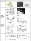
Figure 1
Figure 1
Select biosensor transducer schematics and examples of use. Immunofluorescence schematic [30] and example involving sensing in microgrooves [24]; microcantilevers schematic [6] and example of a fabricated microcantilever array [6]; SPR schematic [22] and example involving detection of concentration of a molecule over time [31]; FRET schematic [32] and example [33] of signal in perturbed and normal cells; impedance schematic [34] and example of signal in control and cells with a toxin [35] (Adapted by permission from Macmillan Publishers Ltd.: J. Invest. Dermatol. [30], ©2013; reused with permission from Elsevier [6,22,24,31,32,34]; with permission ©2008 by the National Academy of Sciences [33]; with permission from Inderscience Ltd. [35], ©Inderscience 2011, respectively).
Figure 2
Figure 2
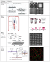
Schematics and examples of selected biofabrication patterning techniques, including microcontact printing [72], inkjet printing [73], MAPLE-DW [74], and LIFT [75,76] (Used with permission from John Wiley and Sons [72]; ©2013 IOP Publishing, reproduced with permission, all rights reserved [73]; used with permission from Elsevier [63,74,75,76], respectively).
Figure 3
Figure 3
Examples of 3D patterned structures with future biosensing application. (a) inkjet printed vascular graft [145], (b) continuous flow printed structure [146], (c) laser patterned cell arrays in hydrogels [144], (d) skin graft fabricated with alternating cell layers [144], (e) laser patterned cell containing microbeads [124], (f) structure of 3D microbead based on an z-stack image of a rhodamine-containing microbead [124], and (g) z-stack image of 3D distribution of cells within an microbead [124] (Used with permission from Elsevier [145]; used with permission from John Wiley and Sons [146]; ©2013 IOP Publishing, reproduced with permission, all rights reserved [124], respectively).

References

    1. Soper S.A., Brown K., Ellington A., Frazier B., Garcia-Manero G., Gau V., Gutman S.I., Hayes D.F., Korte B., Landers J.L., et al. Point-of-care biosensor systems for cancer diagnostics/prognostics. Biosens. Bioelectron. 2006;21:1932–1942. doi: 10.1016/j.bios.2006.01.006. - DOI - PubMed
    1. Tothill I.E. Biosensors for cancer markers diagnosis. Semin. Cell Dev. Biol. 2009;20:55–62. - PubMed
    1. Taitt C.R., Golden J.P., Shubin Y.S., Shriver-Lake L.C., Sapsford K.E., Rasooly A., Ligler F.S. A portable array biosensor for detecting multiple analytes in complex samples. Microb. Ecol. 2004;47:175–185. doi: 10.1007/s00248-003-1011-1. - DOI - PubMed
    1. Curtis T.M., Widder M.W., Brennan L.M., Schwager S.J., van der Schalie W., Fey J., Salazar N. A portable cell-based impedance sensor for toxicity testing of drinking water. Lab Chip. 2009;9:2176–2183. doi: 10.1039/b901314h. - DOI - PubMed
    1. Heeres J.T., Hergenrother P.J. High-throughput screening for modulators of protein-protein interactions: Use of photonic crystal biosensors and complementary technologies. Chem. Soc. Rev. 2011;40:4398–4410. doi: 10.1039/b923660k. - DOI - PubMed