. 2015 Jan 14;10(1):e0116812.
doi: 10.1371/journal.pone.0116812. eCollection 2015.

Detection and quantification of microparticles from different cellular lineages using flow cytometry. Evaluation of the impact of secreted phospholipase A2 on microparticle assessment

Affiliations

Detection and quantification of microparticles from different cellular lineages using flow cytometry. Evaluation of the impact of secreted phospholipase A2 on microparticle assessment

Matthieu Rousseau et al. PLoS One. .

Abstract

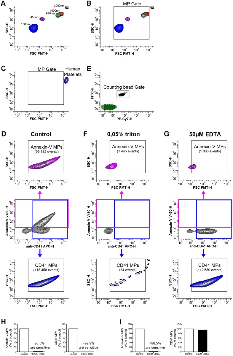
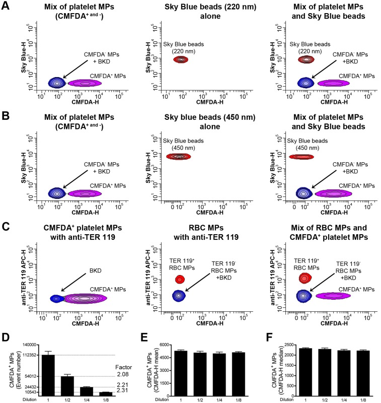
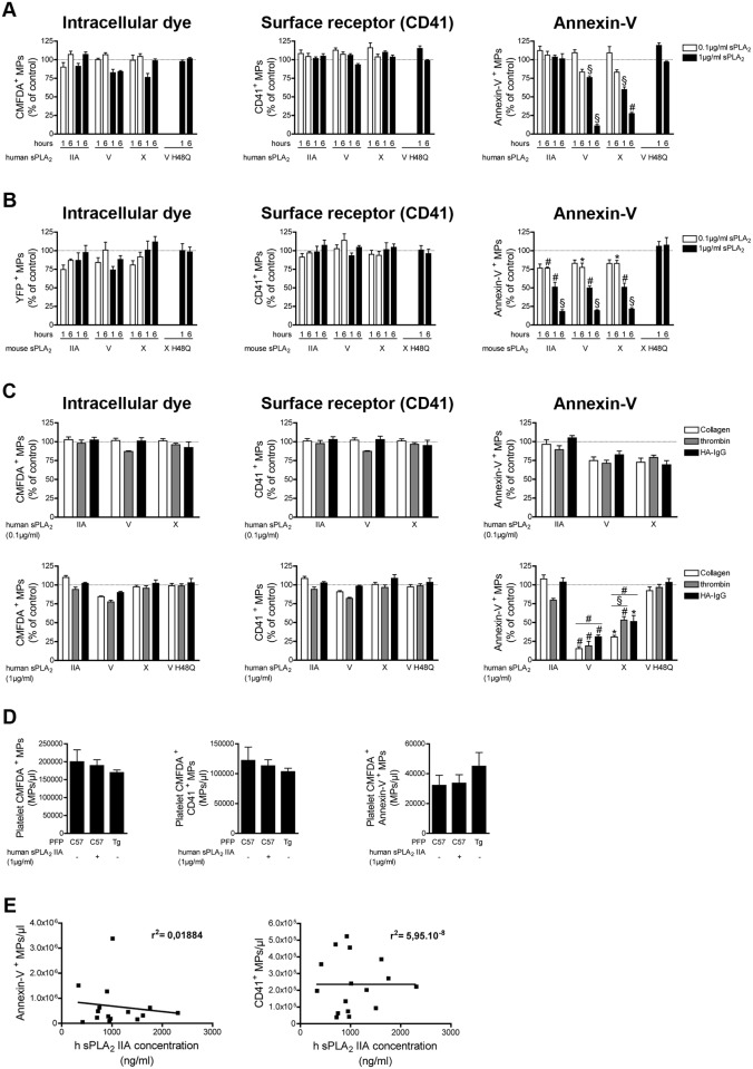
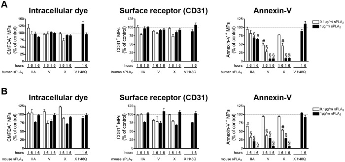
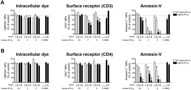
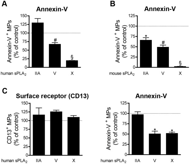
Microparticles, also called microvesicles, are submicron extracellular vesicles produced by plasma membrane budding and shedding recognized as key actors in numerous physio(patho)logical processes. Since they can be released by virtually any cell lineages and are retrieved in biological fluids, microparticles appear as potent biomarkers. However, the small dimensions of microparticles and soluble factors present in body fluids can considerably impede their quantification. Here, flow cytometry with improved methodology for microparticle resolution was used to detect microparticles of human and mouse species generated from platelets, red blood cells, endothelial cells, apoptotic thymocytes and cells from the male reproductive tract. A family of soluble proteins, the secreted phospholipases A2 (sPLA2), comprises enzymes concomitantly expressed with microparticles in biological fluids and that catalyze the hydrolysis of membrane phospholipids. As sPLA2 can hydrolyze phosphatidylserine, a phospholipid frequently used to assess microparticles, and might even clear microparticles, we further considered the impact of relevant sPLA2 enzymes, sPLA2 group IIA, V and X, on microparticle quantification. We observed that if enriched in fluids, certain sPLA2 enzymes impair the quantification of microparticles depending on the species studied, the source of microparticles and the means of detection employed (surface phosphatidylserine or protein antigen detection). This study provides analytical considerations for appropriate interpretation of microparticle cytofluorometric measurements in biological samples containing sPLA2 enzymes.


Conflict of interest statement

Competing Interests: The authors have declared that no competing interests exist.

Figures

Figure 1
Figure 1. Optimization of flow cytometric methods for the detection of MPs.
(A, B) Acquisition of fluorescent microspheres of 100nm (Blue), 450nm (pink), 840nm (green), 1000nm (red), 3200nm (orange) in diameter on a flow cytometer Canto II modified with a FSC-PMT small particles option. (B) A MP gate including particles from 100 to 1000nm in diameter based on the microsphere sizes (FSC-PMT-H) is presented and used to detect MPs. (C) Portrayal of relative size of human platelets detected with fluorochrome-conjugated antibodies directed against CD41. (D) FSC-PMT/SSC portrayal of platelet MPs detected with annexin-V and fluorochrome-conjugated antibodies directed against CD41 in absence of treatment (control). (E) A known concentration of auto-fluorescent polystyrene microspheres (15 µm in diameter) was added in each tube and a determined number of beads was acquired in the counting bead gate to quantitatively process the data. (F, G) FSC-PMT/SSC portrayal of platelet MPs detected with annexin-V and fluorochrome-conjugated antibodies directed against CD41 and treated with 0.05% triton (F) and 50µM EDTA (G). Total annexin-V+ events are detected in the pink gate (middle panel) and the quantity of annexin-V+ MPs is determined in the Annexin-V MP gate (upper panel). Total CD41+ events are detected in the blue gate (middle panel) and the quantity of CD41+ MPs is determined in the CD41 MP gate (lower panel). Data are representative of 5 independent experiments. (H) Triton sensitivity of the platelet MPs detected using fluorochrome-conjugated annexin-V (left panel) and fluorochrome-conjugated antibodies directed against CD41 (right panel) is presented as % of untreated (control). (I) EDTA sensitivity of annexin-V (left panel) and CD41 (right panel) labeling is presented as % of untreated (control). Data are representative of 5 independent experiments.
Figure 2
Figure 2. Study of swarm detection in high sensitivity flow cytometry.
(A) A mixture of CMFDA- and CMFDA+ platelet MPs (CMFDA- and +) and sky blue beads (220 nm in diameter) were analyzed alone (left and middle panel respectively) or mixed (right panel) and their detection resolved on the basis of fluorescence. (B) CMFDA- and + platelet MPs and sky blue beads (450 nm in diameter) were analyzed alone (left and middle panel respectively) or mixed (right panel) prior to detection on the basis of fluorescence. (C) CMFDA+ platelet MPs and RBC MPs labeled with antibodies directed against TER 119 are analyzed alone (left and middle panel respectively) or mixed (right panel). (D, E, F) CMFDA+ platelet MPs were diluted serially thrice (2-fold dilution) and analyzed by high sensitivity flow cytometry to determine their concentration (D), the CMFDA-height (H) mean of fluorescence (E) and the CMFDA-H median of fluorescence (F) are presented. Data are mean ± SEM of 5 independent experiments. BKD = Background noise.
Figure 3
Figure 3. Impact of human and mouse sPLA2s on platelet MPs.
(A) MPs from human platelets (stimulated with collagen) labeled with the CMFDA cell tracker were incubated for 1 and 6 hours at 37°c in absence or in presence of indicated concentrations of human recombinant sPLA2 IIA, V, X, or 1µg/ml of the inactive mutant V H48Q. Fluorochrome-conjugated antibodies directed against CD41 and fluorochrome-conjugated annexin-V were used to assess the quantities of CMFDA+ MPs (left panel), of CD41+ MPs (middle panel), of annexin-V+ MPs (right panel) and were compared to the untreated conditions (dotted line). Data are mean ± SEM of 5 independent experiments presented as % of untreated (control) (B) MPs from mouse platelets (stimulated with collagen), identified using YFP as fluorescent tracker, were incubated 1 and 6 hours at 37°c, in absence or in presence of indicated concentrations of mouse recombinant sPLA2 IIA, V, X, or 1µg/ml of the inactive mutant X H48Q. Fluorochrome-conjugated antibodies directed against CD41 and fluorochrome-conjugated annexin-V were used to determine the concentrations of YFP+ MPs (left panel), of CD41+ MPs (middle panel), of annexin-V+ MPs (right panel) and then compared to the untreated conditions (dotted line). Data are mean ± SEM of 5 independent experiments presented as % of untreated (control). (C) MPs from human platelets labeled with the CMFDA cell tracker and obtained following stimulation with collagen, thrombin or HA-IgG were incubated 6 hours at 37°c in absence or in presence of indicated concentration of human recombinant sPLA2 IIA, V and X and 1µg/ml of the inactive mutant sPLA2 V H48Q. Fluorochrome-conjugated antibodies directed against CD41 and fluorochrome-conjugated annexin-V were used to assess the quantities of CMFDA+ MPs (left panel), of CD41+ MPs (middle panel), of annexin-V+ MPs (right panel) and then compared to the untreated conditions (dotted line). Data are mean ± SEM of 3 independent experiments presented as % of untreated (control). (D) MPs from human platelets (stimulated with collagen) labeled with the CMFDA cell tracker were incubated 6 hours at 37°c in PFP of C57BL6 (supplemented or not with 1µg/ml of recombinant human sPLA2 IIA) or transgenic mice expressing the human sPLA2 IIA (Tg). Fluorochrome-conjugated antibodies directed against CD41 and fluorochrome-conjugated annexin-V were used to assess the quantities of CMFDA+ MPs (left panel), of CMFDA+ CD41+ MPs (middle panel) and CMFDA+ annexin-V+ MPs (right panel). Data are mean ± SEM of 3 independent experiments. (E) Concentrations of Annexin-V+ MPs and CD41+ MPs present in the synovial fluids of RA patients determined by high sensitivity flow cytometry and correlated to the concentration of human sPLA2 IIA assayed (in the same synovial fluids) by time-resolved immunofluorescence analysis. * P< .05; # P< .01; § P< .001.
Figure 4
Figure 4. Impact of human and mouse sPLA2s on erythrocyte MPs.
(A) MPs from human erythrocytes were incubated for 1 and 6 hours at 37°c in absence or in presence of indicated concentrations of human recombinant sPLA2 IIA, V, X, or 1µg/ml of the inactive mutant V H48Q. Fluorochrome-conjugated antibodies directed against CD235a and fluorochrome-conjugated annexin-V were used to assess the quantities of CD235a+ MPs (left panel), of annexin-V+ MPs (right panel) and were compared to the untreated conditions (dotted line). Data are mean ± SEM of 5 independent experiments presented as % of untreated (control) (B) MPs from mouse erythrocytes were incubated 1 and 6 hours at 37°c, in absence or in presence of indicated concentrations of mouse recombinant sPLA2 IIA, V, X, or 1µg/ml of the inactive mutant X H48Q. Fluorochrome-conjugated antibodies directed against TER 119 and fluorochrome-conjugated annexin-V were used to determine the concentrations of TER 119+ MPs (left panel) and annexin-V+ MPs (right panel) and then compared to the untreated conditions (dotted line). Data are mean ± SEM of 5 independent experiments presented as % of untreated (control). * P< .05; # P< .01; § P< .001.
Figure 5
Figure 5. Impact of human and mouse sPLA2s on endothelial cell MPs.
(A) MPs from HUVEC labeled with the CMFDA cell tracker were incubated for 1 and 6 hours at 37°c in absence or in presence of indicated concentrations of human recombinant sPLA2 IIA, V, X, or 1µg/ml of the inactive mutant V H48Q. Fluorochrome-conjugated antibodies directed against CD31 and fluorochrome-conjugated annexin-V were used to assess the quantities of CMFDA+ MPs (left panel), of CD31+ MPs (middle panel), of annexin-V+ MPs (right panel) and were compared to the untreated conditions (dotted line). Data are mean ± SEM of 5 independent experiments presented as % of untreated (control) (B) MPs from mouse EOMA cells labeled with the CMFDA cell tracker were incubated 1 and 6 hours at 37°c, in absence or in presence of indicated concentrations of mouse recombinant sPLA2 IIA, V, X, or 1µg/ml of the inactive mutant X H48Q. Fluorochrome-conjugated antibodies directed against CD31 and fluorochrome-conjugated annexin-V were used to determine the concentrations of CMFDA+ MPs (left panel), of CD31+ MPs (middle panel), of annexin-V+ MPs (right panel) and then compared to the untreated conditions (dotted line). Data are mean ± SEM of 5 independent experiments presented as % of untreated (control). # P< .01; § P< .001.
Figure 6
Figure 6. Impact of sPLA2s on MPs from apoptotic thymocytes.
(A) MPs from human apoptotic thymocytes labeled with the CMFDA cell tracker were incubated for 1 and 6 hours at 37°c in absence or in presence of indicated concentrations of human recombinant sPLA2 IIA, V, X, or 1µg/ml of the inactive mutant V H48Q. Fluorochrome-conjugated antibodies directed against CD3 and fluorochrome-conjugated annexin-V were used to assess the quantities of CMFDA+ MPs (left panel), of CD3+ MPs (middle panel), of annexin-V+ MPs (right panel) and were compared to the untreated conditions (dotted line). Data are mean ± SEM of 5 independent experiments presented as % of untreated (control) (B) MPs from mouse apoptotic thymocytes labeled with the CMFDA cell tracker were incubated 1 and 6 hours at 37°c, in absence or in presence of indicated concentrations of mouse recombinant sPLA2 IIA, V, X, or 1µg/ml of the inactive mutant X H48Q. Fluorochrome-conjugated antibodies directed against CD4 and fluorochrome-conjugated annexin-V were used to determine the concentrations of CMFDA+ MPs (left panel), of CD4+ MPs (middle panel), of annexin-V+ MPs (right panel) and then compared to the untreated conditions (dotted line). Data are mean ± SEM of 5 independent experiments presented as % of untreated (control). * P< .05; # P< .01; § P< .001.
Figure 7
Figure 7. Impact of sPLA2s on MPs from the male reproductive tract.
(A) Human epididymosomes were incubated 6 hours at 37°c in absence or in presence of 1µg/ml of human recombinant sPLA2 IIA, V, X. Fluorochrome-conjugated annexin-V was used to assess the quantities of annexin-V+ MPs and were compared to the untreated conditions (dotted line). Data are mean ± SEM of 3 independent experiments presented as % of untreated (control). (B) Mouse epididymosomes were incubated 6 hours at 37°c in absence or in presence of 1µg/ml of mouse recombinant sPLA2 IIA, V, X. Fluorochrome-conjugated annexin-V was used to assess the quantities of annexin-V+ MPs and were compared to the untreated conditions (dotted line). Data are mean ± SEM of 4 independent experiments presented as % of untreated (control). (C) Human prostasomes were incubated 6 hours at 37°c in absence or in presence of 1µg/ml of human recombinant sPLA2 IIA, V, X. Fluorochrome-conjugated antibodies directed against CD13 and fluorochrome-conjugated annexin-V were used to determine the concentrations of CD13+ MPs (left panel), of annexin-V+ MPs (right panel) and were compared to the untreated conditions (dotted line). Data are mean ± SEM of 4 independent experiments presented as % of untreated (control) * P< .05; # P< .01; § P< .001.

References

    1. Gyorgy B, Szabo TG, Pasztoi M, Pal Z, Misjak P, et al. (2011) Membrane vesicles, current state-of-the-art: emerging role of extracellular vesicles. Cell Mol Life Sci 68: 2667–2688. 10.1007/s00018-011-0689-3 - DOI - PMC - PubMed
    1. Morel O, Jesel L, Freyssinet JM, Toti F (2011) Cellular mechanisms underlying the formation of circulating microparticles. Arterioscler Thromb Vasc Biol 31: 15–26. 10.1161/ATVBAHA.109.200956 - DOI - PubMed
    1. Owens AP 3rd Mackman N (2011) Microparticles in hemostasis and thrombosis. Circ Res 108: 1284–1297. 10.1161/CIRCRESAHA.110.233056 - DOI - PMC - PubMed
    1. Rand ML, Wang H, Bang KW, Packham MA, Freedman J (2006) Rapid clearance of procoagulant platelet-derived microparticles from the circulation of rabbits. J Thromb Haemost 4: 1621–1623. 10.1111/j.1538-7836.2006.02011.x - DOI - PubMed
    1. Rank A, Nieuwland R, Crispin A, Grutzner S, Iberer M, et al. (2011) Clearance of platelet microparticles in vivo. Platelets 22: 111–116. 10.3109/09537104.2010.520373 - DOI - PubMed

Publication types

MeSH terms

Substances