. 2015 Jan 15:12:6.
doi: 10.1186/s12974-014-0221-4.

Differences in the distribution, phenotype and gene expression of subretinal microglia/macrophages in C57BL/6N (Crb1 rd8/rd8) versus C57BL6/J (Crb1 wt/wt) mice

Affiliations

Differences in the distribution, phenotype and gene expression of subretinal microglia/macrophages in C57BL/6N (Crb1 rd8/rd8) versus C57BL6/J (Crb1 wt/wt) mice

Bogale Aredo et al. J Neuroinflammation. .

Abstract

Background: Microglia/macrophages (MG/MΦ) are found in the subretinal space in both mice and humans. Our goal was to study the spatial and temporal distribution, the phenotype, and gene expression of subretinal MG/MΦ in mice with normal retinas and compare them to mice with known retinal pathology.

Methods: We studied C57BL/6 mice with (C57BL/6N), or without (C57BL/6J) the rd8 mutation in the Crb1 gene (which, in the presence of yet unidentified permissive/modifying genes, leads to a retinal degeneration), and documented their fundus appearance and the change with aging. Immunostaining of retinal pigment epithelium (RPE) flat mounts was done for 1) Ionized calcium binding adaptor (Iba)-1, 2) FcγIII/II Receptor (CD16/CD32, abbreviated as CD16), and 3) Macrophage mannose receptor (MMR). Reverse-transcription quantitative PCR (RT-qPCR) was done for genes involved in oxidative stress, complement activation and inflammation.

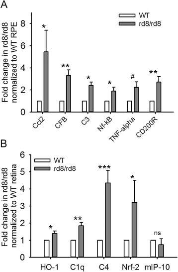
Results: The number of yellow fundus spots correlated highly with subretinal Iba-1+ cells. The total number of subretinal MG/MΦ increased with age in the rd8 mutant mice, but not in the wild-type (WT) mice. There was a centripetal shift in the distribution of the subretinal MG/MΦ with age. Old rd8 mutant mice had a greater number of CD16+ MG/MΦ. CD16+ cells had morphological signs of activation, and this was most prominent in old rd8 mutant mice (P < 1 × 10(-8) versus old WT mice). Subretinal MG/MΦ in rd8 mutant mice also expressed iNOS and MHC-II, and had ultrastructural signs of activation. Finally, rd8 mutant mouse RPE/ MG/MΦ RNA isolates showed an upregulation of Ccl2, CFB, C3, NF-kβ, CD200R and TNF-alpha. The retinas of rd8 mutant mice showed upregulation of HO-1, C1q, C4, and Nrf-2.

Conclusions: When compared to C57BL/6J mice, C57BL/6N mice demonstrate increased accumulation of subretinal MG/MΦ, displaying phenotypical, morphological, and gene-expression characteristics consistent with a pro-inflammatory shift. These changes become more prominent with aging and are likely due to the combination of the rd8 mutation and yet unidentified permissive/modulatory genes in the C57BL/6N mice. In contrast, aging leads to a scavenging phenotype in the C57BL/6J subretinal microglia/macrophages.


Figures
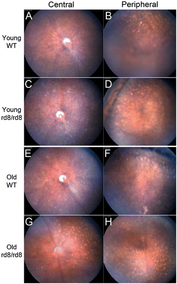
Figure 1
Figure 1
Photographs of central and peripheral retina in C57BL/6N (rd8/rd8) and C57BL/6 J (wild-type) mice. A change in the distribution of fundus spots on B6-mice due to both age and the presence of the rd8 mutation is seen. Yellow spots are shown in central (A, C, E, and G) and peripheral (B, D, F, and H) fundus photographs of representative young (A-D) and old (E-H) B6-mice. Mice 2 to 8 months of age were classified as ‘young’, while mice 14 to 20 months of age were classified as ‘old’. Note that the number of central fundus spots is increased in old age for both rd8 mutant (G versus C) and wild-type (WT) (E versus A) mice. Furthermore, rd8/rd8 mice show a marked increase in central spots compared to WT, both in the young (C versus A) and old (G versus E) age groups.
Figure 2
Figure 2
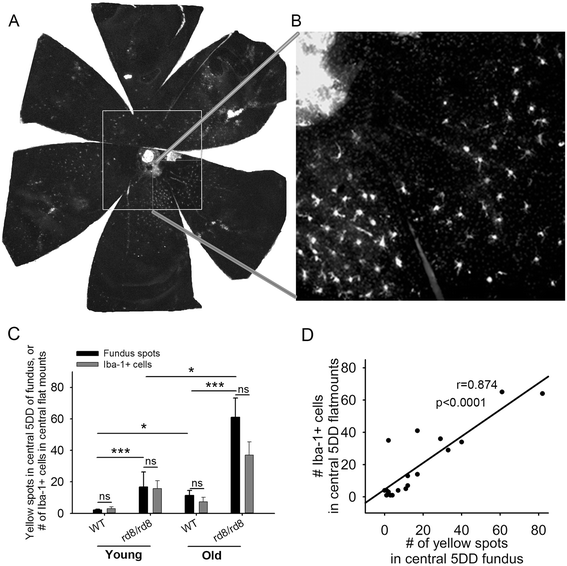
Central fundus spots increase with age in both B6 groups, most prominently in rd8/rd8 mice. (A) A retinal pigment epithelium (RPE) flat mount of an old rd8 mouse stained for ionized calcium binding adaptor (Iba)-1 is shown to demonstrate the area counted as the ‘central flat mount’ (large white square). The ‘central flat mount’ is made up of four higher magnification photos (magnification 10X/1.6X optivar) taken around the disc (smaller squares). (B) One of those higher magnification photos is shown. (C) The number of yellow spots (black bars) in the central fundus (within a circle with a 5 disc diameter (DD) radius and centered on the disc) is very similar to the number of Iba-1+ cells (gray bars) on the corresponding central flat mounts. There is an increase in central fundus spots in rd8/rd8 mice compared to wild-type (WT) mice in each age group. Furthermore, there is an increase in the number of central yellow spots with age in both genotypes (young WT (n = 11), young rd8 (n = 3); old WT (n = 11), old rd8 (n = 10)). (D) Linear regression showing a significant correlation between the number of yellow spots in central fundus (5 DD radius) and the number of Iba-1 positive cells in the corresponding flat mount area (n = 19 eyes). Mice 2 to 8 months of age were classified as ‘young’, while mice 14 to 20 months of age were classified as ‘old’. *P <0.05, ***P <0.001.
Figure 3
Figure 3
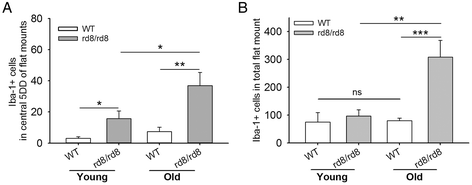
The total number of subretinal microglia/macrophages (MG/MΦ) increases with age in C57BL/6N, but not C57BL/6J mice. (A) Number of ionized calcium binding adaptor (Iba)-1 positive cells in the central flat mount of young and old mice showing a significant increase in both age groups of rd8/rd8 mice compared to the age-matched wild-type (WT). (B) Number of Iba-1 positive cells in the entire/total retinal pigment epithelium (RPE) flat mounts showing that the total number of subretinal microglia increases with age in rd8/rd8 mice, but not in WT mice. The graphs represent the combined results of two to three experiments, which included young mice (WT, n = 10 eyes; or rd8/rd8, n = 8 eyes), and old mice (WT, n = 13 eyes; or rd8/rd8, n = 11 eyes). Mice 2 to 8 months of age were classified as ‘young’, while mice 14 to 20 months of age were classified as ‘old’. *P <0.05, **P <0.01, ***P <0.001.
Figure 4
Figure 4
The phenotype of subretinal microglia/macrophages in B6-mice changes with age and with the rd8 mutation. (A-F) Representative images of triple-stain immunohistochemistry (IHC) show staining with ionized calcium binding adaptor (Iba)-1 (A and D), Macrophage mannose receptor (MMR) (B and E), and FcγIII/II Receptor (CD16/CD32, abbreviated as CD16) (C and F) of subretinal microglia/macrophages (MG/MΦ) on retinal pigment epithelium (RPE)-flat mounts. Different staining patterns are shown here: some Iba-1+ cells stain strongly for CD16 (D and F), while some Iba-1+ cells show weak or no staining for CD16 (A and C). (G) Quantification of MMR + CD16- MG/MΦ in the entire RPE-flat mount shows that in old mice, there is a significant increase in MMR + CD16- cells in rd8/rd8 mice compared to wild-type (WT) mice. (H) In the entire flat mount, there is also a significant increase in the number of CD16+ subretinal MG/MΦ in old rd8/rd8 mice compared to old WT. (I) The central flat mounts demonstrate a significant increase in CD16+ MG/MΦ in old rd8/rd8 mice compared to old WT mice. There is also a trend towards an increase in the number of MMR + CD16- cells in the central flat mounts of old rd8/rd8 mice compared to WT. For figures G-I we combined three similar experiments using 10 young WT, 8 young rd8/rd8, 13 old WT and 10 old rd8/rd8 eyes. Mice 2 to 8 months of age were classified as ‘young’, while mice 14 to 20 months of age were classified as ‘old’. *P <0.05, ***P <0.001, #P = 0.056.
Figure 5
Figure 5
Morphological analysis of subretinal microglia/macrophages in B6-mice. There is an increased microglia/macrophages (MG/MΦ) activation morphology in the rd8/rd8 mice, which is accentuated in old age. (A) The average number of extensions per MG/MΦ cell is decreased in old rd8/rd8 mice compared to both old wild-type (WT) mice and young rd8/rd8 mice. (B) The average length of the MG/MΦ cell extensions (measured using imageJ, http://imageJ.nih.gov/ij/index.html, and expressed as standard arbitrary units) of old rd8/rd8 mice is decreased relative to both old WT mice and young rd8/rd8 mice. (C) Quantification of MG/MΦ activation using the new parameter, microglial morphology activation value (MMAV) is shown. MMAV combines several morphological changes known to be associated with MG/MΦ activation into a single value, and is defined as the area of the MG/MΦ cell body divided by the product of the number of extensions and the average extension length. MMAV is increased in FcγIII/II Receptor (CD16/CD32, abbreviated as CD16) positive cells, particularly in old rd8 mutant mice. (D). The ratio of MMAV for CD16+ to MMAV for CD16- cells is markedly increased in both young and old rd8 mutant mice compared to old WT mice. Two similar experiments were combined (see methods; n = 3 to 5 eyes per group, and 3 to 5 photographic fields per eye, containing 3 to 5 cells with intact cell body per field, which were randomly selected by a masked investigator). Examples of the ionized calcium binding adaptor (Iba)-1 (E,F,I,J) and CD16 (G,H,K,L) staining of MG/MΦ in two C57BL/6J (E,G,I,K) versus two C57BL/6N mice (F,H,J,L) are shown. Mice 2 to 8 months of age were classified as ‘young’, while mice 14 to 20 months of age were classified as ‘old’. *P <0.05, **P <0.01, ***P <0.001, #P = 0.051.
Figure 6
Figure 6
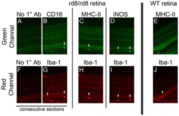
Subretinal microglia/macrophages (MG/MΦ) in C57BL/6N mice express markers of activation. After processing eyes using the freeze-substitution technique (see Methods, n = 8 eyes per group), retina sections from C57BL/6N (A-D and F-I) or from C57BL/6J (E and J) eyes were obtained and stained. Each section was double-labeled with ionized calcium binding adaptor (Iba)-1 (red channel; G-J) and one microglia/macrophage activation marker (green channel; B-E). A control sample is also shown (secondary antibodies without primary antibodies; A and F), which is a consecutive section of the one stained with Iba-1 and FcγIII/II Receptor (CD16/CD32, abbreviated as CD16) (B and G). Note the expression of CD16 and activation markers MHC-II and iNOS in subretinal MG/MΦ in rd8 mutant samples (arrows in B,C,D). There is no staining for MHC-II in the wild-type (WT) subretinal MG/MΦ (E versus C).
Figure 7
Figure 7
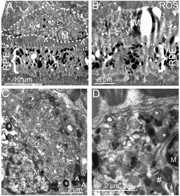
Electron microscopy of retinal sections in old rd8/rd8 mice. Cells consistent with microglia/macrophages (MG/MΦ) were seen between the retinal pigment epithelium (RPE) and the photoreceptor outer segments (A,B). Higher magnification images from these cells (C,D) demonstrate the presence of photoreceptor outer segment debris (labeled with ^), occasional melanosomes, lipofuscin granules (*), and multiple phagosomes with partially degraded debris (#). The photoreceptor outer segments seen over the microglia in A and B are labeled as ROS (rod outer segments).
Figure 8
Figure 8
RT-qPCR analysis of RNA from retinal pigment epithelium (RPE)/subretinal microglia/macrophages (MG/MΦ) isolates and from retina isolates. There is an upregulation of genes related to oxidative stress, complement activation and inflammation in rd8 mutant mice compared to wild-type (WT). (A) Fold changes in the expression of target genes, in RNA isolated from RPE/subretinal MG/MΦ from rd8 mutant mice, normalized to B6-WT. The bars represent the average of two experiments in age-matched 6- to 8-month-old mice, including a total of 10 rd8/rd8 and 10 WT eyes, except for CFB (6 rd8/rd8 and 6 WT eyes). (B) Fold changes in the expression of target genes in RNA isolated from the retina of rd8 mutant mice normalized to B6-WT. The bars represent the average of two experiments including a total of 11 rd8/rd8 and 11 B6 eyes. *P <0.05, **P <0.01, ***P <0.001, #P = 0.058.

References

    1. Mrak RE. Microglia in Alzheimer brain: a neuropathological perspective. Int J Alzheimers Dis. 2012;2012:165021. - PMC - PubMed
    1. Ridolfi E, Barone C, Scarpini E, Galimberti D. The role of the innate immune system in Alzheimer’s disease and frontotemporal lobar degeneration: an eye on microglia. Clin Dev Immunol. 2013;2013:939786. doi: 10.1155/2013/939786. - DOI - PMC - PubMed
    1. Li Y, Liu L, Liu D, Woodward S, Barger SW, Mrak RE, Griffin WS. Microglial activation by uptake of fDNA via a scavenger receptor. J Neuroimmunol. 2004;147:50–5. doi: 10.1016/j.jneuroim.2003.10.043. - DOI - PMC - PubMed
    1. Penfold PL, Killingsworth MC, Sarks SH. Senile macular degeneration. The involvement of giant cells in atrophy of the retinal pigment epithelium. Invest Ophthalmol Vis Sci. 1986;27:364–71. - PubMed
    1. Penfold PL, Killingsworth MC, Sarks SH. Senile macular degeneration: the involvement of immunocompetent cells. Graefes Arch Clin Exp Ophthalmol. 1985;223:69–76. doi: 10.1007/BF02150948. - DOI - PubMed

Publication types

MeSH terms