. 2015 Mar;92(3):68.
doi: 10.1095/biolreprod.114.122887. Epub 2015 Jan 14.

Progesterone-based intrauterine device use is associated with a thinner apical layer of the human ectocervical epithelium and a lower ZO-1 mRNA expression

Affiliations

Progesterone-based intrauterine device use is associated with a thinner apical layer of the human ectocervical epithelium and a lower ZO-1 mRNA expression

Annelie Tjernlund et al. Biol Reprod. 2015 Mar.

Abstract

Currently, whether hormonal contraceptives affect male to female human immunodeficiency virus (HIV) transmission is being debated. In this study, we investigated whether the use of progesterone-based intrauterine devices (pIUDs) is associated with a thinning effect on the ectocervical squamous epithelium, down-regulation of epithelial junction proteins, and/or alteration of HIV target cell distribution in the human ectocervix. Ectocervical tissue biopsies from healthy premenopausal volunteers using pIUDs were collected and compared to biopsies obtained from two control groups, namely women using combined oral contraceptives (COCs) or who do not use hormonal contraceptives. In situ staining and image analysis were used to measure epithelial thickness and the presence of HIV receptors in tissue biopsies. Messenger RNA levels of epithelial junction markers were measured by quantitative PCR. The epithelial thickness displayed by women in the pIUD group was similar to those in the COC group, but significantly thinner as compared to women in the no hormonal contraceptive group. The thinner epithelial layer of the pIUD group was specific to the apical layer of the ectocervix. Furthermore, the pIUD group expressed significantly lower levels of the tight junction marker ZO-1 within the epithelium as compared to the COC group. Similar expression levels of HIV receptors and coreceptors CD4, CCR5, DC-SIGN, and Langerin were observed in the three study groups. Thus, women using pIUD displayed a thinner apical layer of the ectocervical epithelium and reduced ZO-1 expression as compared to control groups. These data suggest that pIUD use may weaken the ectocervical epithelial barrier against invading pathogens, including HIV.

Keywords: HIV receptors; epithelial junction proteins; female reproductive tract; hormonal contraception; in situ staining.


Figures

FIG. 1
FIG. 1
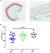
Users of pIUDs display a thinner ectocervical epithelium. A) Bright field images of ectocervical tissue sections showing how the height of the epithelium was measured by drawing lines from the basal membrane to the top/last cell layer of the epithelium. The height measurements were taken in intervals of approximately 100 μm along the entire length of the epithelium. The image on the left was collected with a 2.5× objective while the image on the right was captured with a 20× objective. B) Graphs showing the distribution and median of epithelial thickness (μm) in the three study groups. For the noHC group, empty symbols represent women in the early stage of the menstrual cycle and filled symbols represent women in the late stage of the menstrual cycle. A nonparametric, two-tailed Mann-Whitney U-test was used to compare pIUD versus COC and pIUD versus noHC; P < 0.05 was considered statistically significant.
FIG. 2
FIG. 2
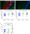
The apical layer of the ectocervical epithelium is thinner in pIUD users. A) Immunofluorescence images of ectocervical tissue sections stained for the adhesion junction protein E-cadherin (red) (left image) or stained with IgG isotype control (right image) was used to demonstrate the localization of the different layers of ectocervical epithelium. 4′,6-Diamidino-2-phenylindole (blue) was used as a counterstain for visualization of cell nuclei. The total height of the ectocervical epithelium was measured from the basal membrane (BM; indicated by the blue arrow) to the last cell layer of the ectocervical epithelium (indicated by the green arrow). The height of the stratum malpighii (SM) was measured from the basal membrane to the end of the E-cadherin-expressing layer (indicated by the red arrow). The stratum corneum (SC) was measured from the E-cadherin-expressing layer to the last cell layer of the ectocervical epithelium. The images were collected with a 40× objective. Bar = 40 μm. The graphs show the distribution and median of the total epithelial thickness (B), the thickness of the stratum malpighii (C), and the thickness of the stratum corneum (D) in the three study groups. For the noHC group, empty symbols represent women in the early stage of the menstrual cycle and filled symbols represent women in the late stage of the menstrual cycle. A nonparametric, two-tailed Mann-Whitney U-test was used to compare pIUD versus COC and pIUD versus noHC; P < 0.05 was considered statistically significant.
FIG. 3
FIG. 3
Use of pIUDs lowers the level of ZO-1 mRNA. Quantitative RT-PCR was performed to determine the distribution and median of relative quantification (RQ) of the mRNA expression levels of the epithelial junction markers E-cadherin (A), ZO-1 (B), claudin-1 (C), and occludin (D). The cycle threshold (Ct) values for the selected markers were normalized to UBC by using the 2−dCT method. The two outliers exhibited undetectable ZO-1 expression level and were assigned a reference value above Ct 40. For the noHC group, empty symbols represent women in the early stage of the menstrual cycle and filled symbols represent women in the late stage of the menstrual cycle. A nonparametric, two-tailed Mann-Whitney U-test was used to compare pIUD versus COC and pIUD versus noHC; P < 0.05 was considered statistically significant.
FIG. 4
FIG. 4
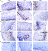
Similar expression of HIV receptor in the ectocervical epithelium in the three study groups. Bright field images of ectocervical tissue sections showing the in situ expression of HIV receptors. The tissue sections were stained with hematoxylin (blue) for visualization of cell nuclei and stained brown for CD4 (A), CCR5 (B), DC-SIGN (C), Langerin (D), and IgG isotype control (E). The images in the first, middle, and right columns represent the pIUD, COC, and noHC groups, respectively. The negative controls did not contain any positively stained cells. The images were collected with a 40× objective. Bar = 100 μm.
FIG. 5
FIG. 5
Similar frequencies of HIV receptor expression in the ectocervical epithelium in the three study groups. The graphs show the distribution and median of the percentage of CD4+ (A), CCR5+ (B), DC-SIGN+ (C), and Langerin+ (D) cells relative to the total epithelial tissue analyzed. For the noHC group, empty symbols represent women in the early stage of the menstrual cycle and filled symbols represent women in the late stage of the menstrual cycle. Note that one data point is missing for the frequency of CD4+ and CCR5+ cells in the noHC group due to technical difficulties. A nonparametric, two-tailed Mann-Whitney U-test was used to analyze statistical significance between the study groups; P < 0.05 was considered statistically significant.

References

    1. World Contraceptive Patterns 2013. [Internet] Population Division, United Nations Department of Economic and Social Affairs; http://www.un.org/en/development/desa/population/publications/family/con.... Accessed 1 June 2014.
    1. Blish CA, Baeten JM. Hormonal contraception and HIV-1 transmission. Am J Reprod Immunol. 2011;65:302–307. - PMC - PubMed
    1. Chandra N, Thurman AR, Anderson S, Cunningham TD, Yousefieh N, Mauck C, Doncel GF. Depot medroxyprogesterone acetate increases immune cell numbers and activation markers in human vaginal mucosal tissues. AIDS Res Hum Retroviruses. 2013;29:592–601. - PMC - PubMed
    1. Critchlow CW, Wolner-Hanssen P, Eschenbach DA, Kiviat NB, Koutsky LA, Stevens CE, Holmes KK. Determinants of cervical ectopia and of cervicitis: age, oral contraception, specific cervical infection, smoking, and douching. Am J Obstet Gynecol. 1995;173:534–543. - PubMed
    1. Wira CR, Fahey JV, Rodriguez-Garcia M, Shen Z, Patel MV. Regulation of mucosal immunity in the female reproductive tract: the role of sex hormones in immune protection against sexually transmitted pathogens. Am J Reprod Immunol. 2014;72:236–258. - PMC - PubMed

Publication types

MeSH terms