. 2015 Feb 15;128(4):755-67.
doi: 10.1242/jcs.161786. Epub 2015 Jan 14.

ESCRT-0 marks an APPL1-independent transit route for EGFR between the cell surface and the EEA1-positive early endosome

Affiliations

ESCRT-0 marks an APPL1-independent transit route for EGFR between the cell surface and the EEA1-positive early endosome

Neftali Flores-Rodriguez et al. J Cell Sci. .

Abstract

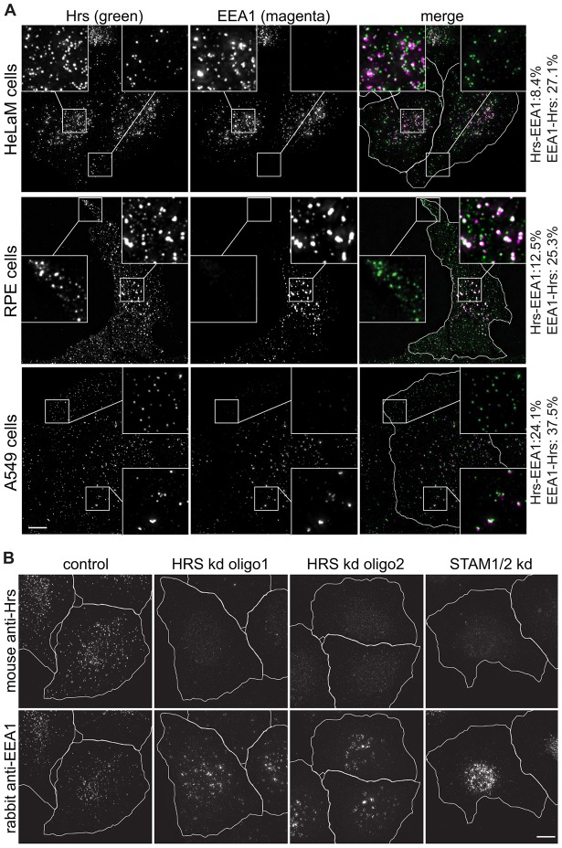
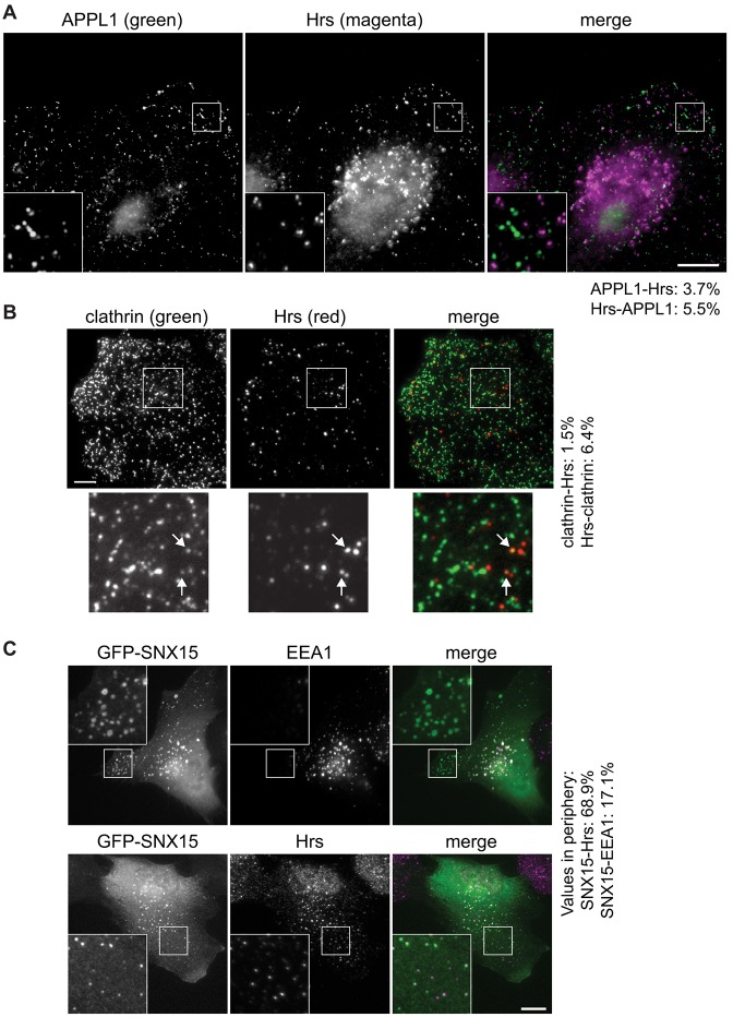
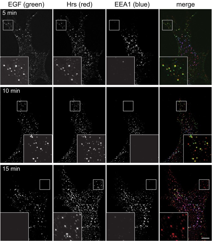
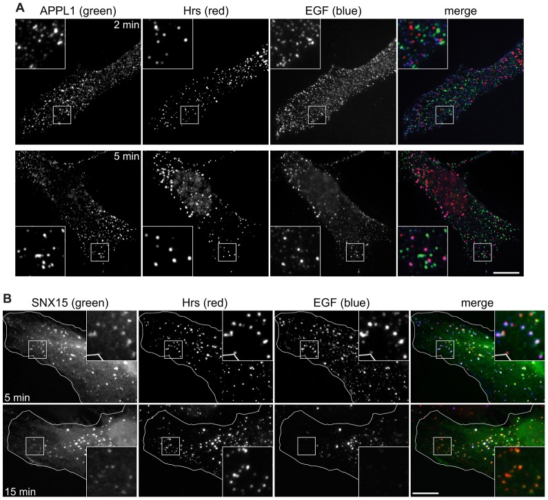
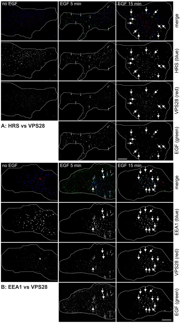
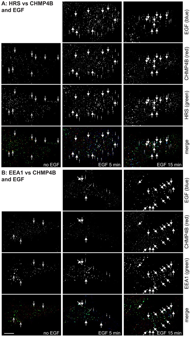
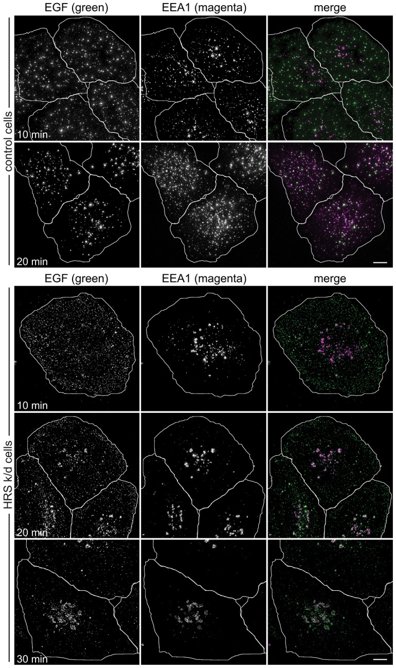
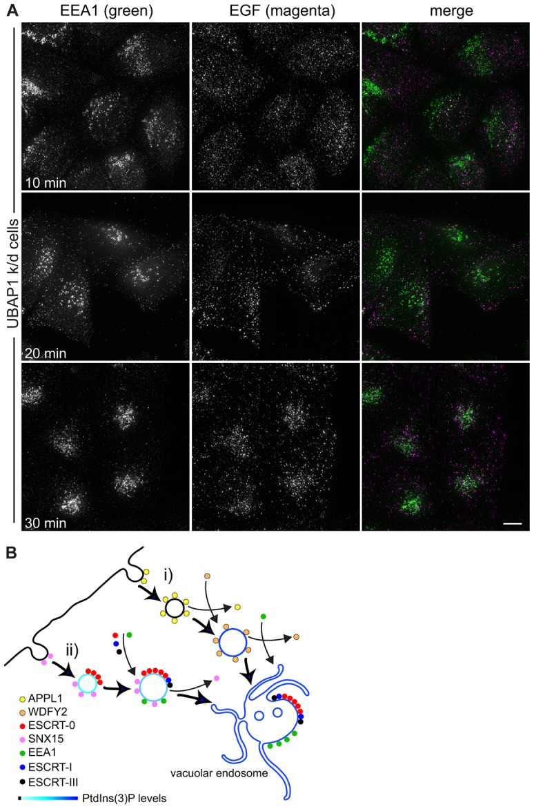
Endosomal sorting complexes required for transport (ESCRT)-0 sorts ubiquitylated EGFR within the early endosome so that the receptor can be incorporated into intralumenal vesicles. An important question is whether ESCRT-0 acts solely upon EGFR that has already entered the vacuolar early endosome (characterised by the presence of EEA1) or engages EGFR within earlier compartments. Here, we employ a suite of software to determine the localisation of ESCRT-0 at subpixel resolution and to perform particle-based colocalisation analysis with other endocytic markers. We demonstrate that although some of the ESCRT-0 subunit Hrs (also known as HGS) colocalises with the vacuolar early endosome marker EEA1, most localises to a population of peripheral EEA1-negative endosomes that act as intermediates in transporting EGFR from the cell surface to more central early endosomes. The peripheral Hrs-labelled endosomes are distinct from APPL1-containing endosomes, but co-label with the novel endocytic adaptor SNX15. In contrast to ESCRT-0, ESCRT-I is recruited to EGF-containing endosomes at later times as they move to more a central position, whereas ESCRT-III is also recruited more gradually. RNA silencing experiments show that both ESCRT-0 and ESCRT-I are important for the transit of EGF to EEA1 endosomes.

Keywords: EEA1; EGFR; ESCRT-I; Hrs; Particle-based colocalisation.


Figures

Fig. 1.
Fig. 1.
Identification of peripheral Hrs-positive endosomes lacking EEA1. (A) HeLaM (upper panel), RPE (centre panel) or A549 cells (lower panel) were co-stained with rabbit anti-Hrs and mouse anti-EEA1. (B) HeLaM cells were knocked down for ESCRT-0 components as indicated and stained with mouse anti-Hrs and rabbit anti-EEA1. The insets show a ×3 magnification of the indicated areas. Values show net percentages (i.e. scrambled values subtracted) of particles labelled by marker 1 that are also labelled by marker 2. kd, knockdown. Cell outlines are shown in white. Scale bars: 10 µm.
Fig. 2.
Fig. 2.
Localisation of peripheral Hrs to SNX15 but not APPL1 endosomes. (A) HeLaM cells were co-stained for APPL1 and Hrs, and were examined by wide-field microscopy. (B) HeLaM cells were stained for clathrin and Hrs and imaged by TIRF microscopy. Arrows show examples of structures containing both Hrs and clathrin. (C) HeLaM cells were transfected with GFP–SNX15, fixed and stained for EEA1 and Hrs and examined by wide-field microscopy. The insets show a ×3 magnification of the indicated areas. Values show net percentages (i.e. scrambled values subtracted) of particles labelled by marker 1 that are also labelled by marker 2. Scale bars: 10 µm.
Fig. 3.
Fig. 3.
EGF passes through peripheral Hrs endosomes prior to labelling EEA1 endosomes. RPE cells were serum starved, then pulsed for 2 min with fluorescent EGF, then chased for a further 3 min (upper row), 8 min (middle row) or 13 min (lower row) before fixing and labelling with antibodies against Hrs or EEA1. The insets show a ×3 magnification of the indicated areas. Scale bar: 10 µm.
Fig. 4.
Fig. 4.
EGF labels distinct Hrs/SNX15-positive and APPL1-positive endosomes. Untransfected RPE cells (A) or RPE cells transfected with GFP–SNX15 (B) were serum starved and then pulse-chased with fluorescent EGF for the indicated total times and labelled for immunofluorescence. Cell outlines are shown in white. The insets show a ×3 magnification of the indicated areas. Scale bars: 10 µm.
Fig. 5.
Fig. 5.
Localisation of ESCRT-I to early endosomes. RPE cells were serum starved and pulse-chased with fluorescent EGF for the indicated total times, permeabilised with saponin and labelled for VPS28 and Hrs (A) or EEA1 (B). Small arrows show examples of endosomes containing EGF and Hrs or EEA1 but lacking VPS28 labelling. Large arrows highlight example endosomes containing all three markers. Cell outlines are shown in white. Scale bars: 10 µm.
Fig. 6.
Fig. 6.
Localisation of ESCRT-III to early endosomes. RPE cells were serum starved and pulse-chased with fluorescent EGF for the indicated total times, permeabilised with saponin and labelled for CHMP4B and Hrs (A) or EEA1 (B). Arrows with open arrowheads show examples of CHMP4B in unstimulated cells colocalising with Hrs or EEA1. Small arrows show examples of endosomes containing EGF and Hrs or EEA1 but lacking CHMP4B labelling. Large arrows highlight example endosomes containing all three markers. Scale bar: 10 µm.
Fig. 7.
Fig. 7.
ESCRT-0 is required for EGF transit to EEA1 endosomes. HeLaM cells transfected with control siRNA or Hrs siRNA oligo 1 were pulse-chased with fluorescent EGF for the indicated total times, fixed for immunofluorescence and labelled with anti-EEA1. Cell outlines are shown in white. k/d, knockdown. Scale bars: 10 µm.
Fig. 8.
Fig. 8.
ESCRTs and the organisation of the early endocytic pathway. (A) HeLaM cells transfected with UBAP1 siRNA oligo 1 were pulse-chased with fluorescent EGF for the indicated total times and fixed for immunofluorescence. Scale bar: 10 µm. (B) EGFR can enter the endocytic system through at least two clathrin-mediated pathways. (i) APPL1 associates with clathrin pits and peripheral vesicles, then is replaced by WDFY2 as PtdIns3P is generated. WDFY2 in turn is replaced by EEA1 (see Zoncu et al., 2009). (ii) SNX15 and ESCRT-0 bind to a separate population of early endocytic intermediates. Generation of increasing levels of PtdIns3P as the vesicle develops, combined with increasing ubiquitylation of EGFR, might recruit more ESCRT-0, and ESCRT-I and ESCRT-III are also recruited alongside EEA1 as the endosome matures. ESCRT-0, but not SNX15 (Danson et al., 2013), enters clathrin-rich domains on the vacuolar endosome.

References

    1. Ali N., Zhang L., Taylor S., Mironov A., Urbé S., Woodman P. (2013). Recruitment of UBPY and ESCRT exchange drive HD-PTP-dependent sorting of EGFR to the MVB. Curr. Biol. 23, 453–461 10.1016/j.cub.2013.02.033 - DOI - PubMed
    1. Babst M., Wendland B., Estepa E. J., Emr S. D. (1998). The Vps4p AAA ATPase regulates membrane association of a Vps protein complex required for normal endosome function. EMBO J. 17, 2982–2993 10.1093/emboj/17.11.2982 - DOI - PMC - PubMed
    1. Bache K. G., Slagsvold T., Cabezas A., Rosendal K. R., Raiborg C., Stenmark H. (2004). The growth-regulatory protein HCRP1/hVps37A is a subunit of mammalian ESCRT-I and mediates receptor down-regulation. Mol. Biol. Cell 15, 4337–4346 10.1091/mbc.E04-03-0250 - DOI - PMC - PubMed
    1. Barysch S. V., Aggarwal S., Jahn R., Rizzoli S. O. (2009). Sorting in early endosomes reveals connections to docking- and fusion-associated factors. Proc. Natl. Acad. Sci. USA 106, 9697–9702 10.1073/pnas.0901444106 - DOI - PMC - PubMed
    1. Bishop N., Woodman P. (2001). TSG101/mammalian VPS23 and mammalian VPS28 interact directly and are recruited to VPS4-induced endosomes. J. Biol. Chem. 276, 11735–11742 10.1074/jbc.M009863200 - DOI - PubMed

Publication types

MeSH terms