Review
. 2015 Jan;5(1):327-96.
doi: 10.1002/cphy.c130056.

Neural control of the lower urinary tract

Affiliations
Review

Neural control of the lower urinary tract

William C de Groat et al. Compr Physiol. 2015 Jan.

Abstract

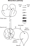
This article summarizes anatomical, neurophysiological, pharmacological, and brain imaging studies in humans and animals that have provided insights into the neural circuitry and neurotransmitter mechanisms controlling the lower urinary tract. The functions of the lower urinary tract to store and periodically eliminate urine are regulated by a complex neural control system in the brain, spinal cord, and peripheral autonomic ganglia that coordinates the activity of smooth and striated muscles of the bladder and urethral outlet. The neural control of micturition is organized as a hierarchical system in which spinal storage mechanisms are in turn regulated by circuitry in the rostral brain stem that initiates reflex voiding. Input from the forebrain triggers voluntary voiding by modulating the brain stem circuitry. Many neural circuits controlling the lower urinary tract exhibit switch-like patterns of activity that turn on and off in an all-or-none manner. The major component of the micturition switching circuit is a spinobulbospinal parasympathetic reflex pathway that has essential connections in the periaqueductal gray and pontine micturition center. A computer model of this circuit that mimics the switching functions of the bladder and urethra at the onset of micturition is described. Micturition occurs involuntarily in infants and young children until the age of 3 to 5 years, after which it is regulated voluntarily. Diseases or injuries of the nervous system in adults can cause the re-emergence of involuntary micturition, leading to urinary incontinence. Neuroplasticity underlying these developmental and pathological changes in voiding function is discussed.


Figures

Figure 1
Figure 1
Efferent pathways of the lower urinary tract. (A) Innervation of the female lower urinary tract. Sympathetic fibers (shown in blue) originate in the T11-L2 segments in the spinal cord and run through the inferior mesenteric plexus (IMP) and the hypogastric nerve (HGN) or through the paravertebral chain to enter the pelvic nerves at the base of the bladder and the urethra. Parasympathetic preganglionic fibers (shown in green) arise from the S2–S4 spinal segments and travel in sacral roots and pelvic nerves (PEL) to ganglia in the pelvic plexus (PP) and in the bladder wall. This is where the postganglionic nerves that supply parasympathetic innervation to the bladder arise. Somatic motor nerves (shown in yellow) that supply the striated muscles of the external urethral sphincter arise from S2–S4 motor neurons and pass through the pudendal nerves. (B) Efferent pathways and neurotransmitter mechanisms that regulate the lower urinary tract. Parasympathetic postganglionic axons in the pelvic nerve release acetylcholine (ACh), which produces a bladder contraction by stimulating M3 muscarinic receptors in the bladder smooth muscle. Sympathetic postganglionic neurons release noradrenaline (NA), which activates β3 adrenergic receptors to relax bladder smooth muscle and activates α1 adrenergic receptors to contract urethral smooth muscle. Somatic axons in the pudendal nerve also release ACh, which produces a contraction of the external sphincter striated muscle by activating nicotinic cholinergic receptors. Parasympathetic postganglionic nerves also release ATP, which excites bladder smooth muscle, and nitric oxide, which relaxes urethral smooth muscle (not shown). L1, first lumbar root; S1, first sacral root; SHP, superior hypogastric plexus; SN, sciatic nerve; T9, ninth thoracic root (216).
Figure 2
Figure 2
Classes and distribution of afferent nerves in the LUT. (A) The distribution of the different classes of fibers in the bladder wall and urethra. (B) In the pelvic nerve, four types of mechanosensitive fibers were identified by stretch, stroke, and probe. (C) Proportions of afferent fiber types recorded in the pelvic nerve. (D) Distribution of low- and high-threshold receptive fields of pelvic nerve muscle afferent fibers based on responses to stretch. (E) Distribution of receptive fields of the four classes of pelvic nerve fibers (303).
Figure 3
Figure 3
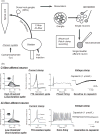
(A) Experimental methods for performing patch-clamp recordings on bladder afferent neurons obtained from rats with chronic cystitis. Chronic cystitis was induced by intraperitoneal injection of cyclophosphamide. Fluorescent dye (fast blue) injected into the bladder wall was transported via Aδ- and C-fiber bladder afferent axons to neurons in the dorsal root ganglia (DRG). L6 and S1 DRG were dissected and dissociated into single neurons by enzymatic methods. Whole cell patch-clamp recordings were then performed on fast blue-labeled bladder afferent neurons that were identified with a fluorescence microscope. (B) Characteristics of a bladder afferent neuron (24-μm diameter, C-fiber afferent neuron, top record) exhibiting tetrodotoxin (TTX)-resistant action potentials and a bladder afferent neuron (33-μm diameter, Aδ-fiber afferent neuron, bottom record) exhibiting TTX-sensitive action potentials. The left panels are voltage responses and action potentials evoked by 30-ms depolarizing current pulses injected through the patch pipette in current-clamp conditions. Asterisks with dashed lines indicate the thresholds for spike activation. The second panels on the left side show the effects of TTX application (1 μmol/L) on action potentials. The third panels from the left show firing patterns during membrane depolarization (700-ms duration). The panels on the right show the responses to extracellular application of capsaicin (1 μmol/L) in voltage-clamp conditions. Note that the C-fiber afferent neuron exhibited TTX-resistant phasic firing (i.e., one to two spikes during prolonged membrane depolarization) and an inward current in response to capsaicin, while A-fiber afferent neuron exhibited TTX-sensitive tonic firing (i.e., repetitive firing during membrane depolarization) and no response to capsaicin (176).
Figure 4
Figure 4
Hypothetical model depicting possible interactions between bladder afferent nerves, urothelial cells, smooth muscle cells, interstitial cells, and blood vessels. Urothelial cells can also be targets for transmitters released from nerves or other cell types. Urothelial cells can be activated by either autocrine (i.e., autoregulation) or paracrine mechanisms (release from nearby nerves or other cells). Bladder stretch releases ATP which acts on P2 receptors on the afferent terminal or the interstitial cell and on P2 receptors on the urothelial cell. Stretch also releases ACh which acts on muscarinic receptors (M3) on the afferent terminal, the interstial cell or the urothelial cell. The latter action can release NO. Epinephrine or norepinephrine also release NO from the urothelial cell by activating β3 adrenergic receptors (50).
Figure 5
Figure 5
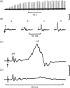
Extracellular recordings on a bladder postganglionic nerve demonstrating facilitation of pelvic ganglionic transmission during repetitive stimulation of preganglionic axons in the pelvic nerve. (A) Action potentials evoked with submaximal (5V, 1 Hz) stimulation recorded with a slow time base. (B) Sample responses from A (a, 1st; b, 5th; c, 10th and d, 20th responses in the series) obtained, respectively, 5, 10, and 20 s later and recorded with a fast time base. (C) Average (20 sweeps) of a facilitated ganglionic response (I.I V, I Hz) showing the nonsynaptic, early response (ER) consisting of axonal volley and the facilitated late synaptic response (LR). (D) Depression by hexamethonium (100 μg, I.A.) of the late (synaptic) response but no effect on the early (nonsynaptic) response. The arrow below C denotes the stimulus artifact. Time calibration in D also applies to C; vertical calibration in A and B is 400 μV and in C and D is 200 μV, negativity upward (171).
Figure 6
Figure 6
(A) Diagram showing the autonomic innervation of the urinary bladder of the cat and the synaptic mechanisms within bladder ganglia. Nicotinic (N), muscarinic (M), and adrenergic (A) receptors are depicted on a principal ganglion cell and a small intensely fluorescent cell (SIF cell). Receptors mediating hyperpolarization (H), depolarization (D) are also indicated. An α-adrenergic receptor (A, α-INH) mediating presynaptic inhibition is indicated on the preganglionic nerve terminal. Inhibitory and excitatory synaptic mechanisms are designated by − and +, respectively. Postsynaptic adrenergic and muscarinic receptors mediate both hyperpolarizing and depolarizing responses. Sympathetic preganglionic axons make synaptic contact with cells in the inferior mesenteric ganglion (IMG) and also send axons through the IMG to make synaptic contacts with SIF cells in bladder ganglia. SIF cells have both nicotinic and muscarinic excitatory receptors. Sympathetic efferent pathways can be activated by afferent projections from the urinary bladder. (B) Suppression of EPSP amplitude by exogenous norepinephrine (NE, 3 × 10⁻⁴). Membrane potential (0) and EPSP amplitude (●) recorded during preganglionic stimulation and following application of NE, (bar). (C) Series of 10 superimposed sweeps showing fast excitatory postsynaptic potentials (f-EPSPs) and action potentials (truncated) elicited before (#1), after start (#2 and #3) of perfusion with NE, (1 × 10⁻⁴) and 1 (#4), 8 (#5), and 16 min (#6) following return to the control solution. NE depressed f-EPSP amplitude and spike generation (151).
Figure 7
Figure 7
Reflex voiding responses in an infant, a healthy adult, and a paraplegic patient. Combined cystometrograms and sphincter electromyograms (EMGs, recorded with surface electrodes), allowing a schematic comparison of reflex voiding responses in an infant (A) and in a paraplegic patient (C) with a voluntary voiding response in a healthy adult (B). The abscissa in all recordings represents bladder volume in millilitres; the ordinates represent electrical activity of the EMG recording and detrusor pressure (the component of bladder pressure that is generated by the bladder itself) in cmH2O. On the left side of each trace (at 0 mL), a slow infusion of fluid into the bladder is started (bladder filling). In part B, the start of sphincter relaxation, which precedes the bladder contraction by a few seconds, is indicated (“start”). Note that a voluntary cessation of voiding (“stop”) is associated with an initial increase in sphincter EMG and detrusor pressure (a myogenic response). A resumption of voiding is associated with sphincter relaxation and a decrease in detrusor pressure that continues as the bladder empties and relaxes. In the infant (A), sphincter relaxation is present but less complete. On the other hand, in the paraplegic patient (C) the reciprocal relationship between bladder and sphincter is abolished. During bladder filling, involuntary bladder contractions (detrusor overactivity) occur in association with sphincter activity. Each wave of bladder contraction is accompanied by simultaneous contraction of the sphincter (detrusor-sphincter dyssynergia), hindering urine flow. Loss of the reciprocal relationship between the bladder and the sphincter in paraplegic patients thus interferes with bladder emptying (216).
Figure 8
Figure 8
A simple working model of the lower urinary tract control system, showing the voiding reflex and brainstem (green) and circuits 1, 2, and 3 (red/blue, yellow, and blue, respectively). PAG = periaqueductal gray; PMC = pontine micturition center; th = thalamus; mPFC = medial prefrontal cortex; lPFC = lateral prefrontal cortex; SMA = supplementary motor area; dACC = dorsal anterior cingulate cortex.
Figure 9
Figure 9
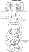
Neural connections between the brain and the sacral spinal cord that may regulate the lower urinary tract in the cat. Lower section of the sacral spinal cord shows the location and morphology of a preganglionic neuron in the sacral parasympathetic nucleus (PSN), a sphincter motor neuron in Onuf’s nucleus (ON), and the sites of central termination of afferent projections (shaded area) from the urinary bladder. Upper section of the sacral cord shows the sites of termination (shaded areas) of descending pathways arising from the medial pontine micturition center (PMC), the lateral pontine sphincter or urine storage center, and the paraventricular nuclei of the hypothalamus. Section through the pons shows the projection from the anterior hypothalamic nuclei to the pontine micturition center (PMC).
Figure 10
Figure 10
Primary afferent and spinal interneuronal pathways involved in micturition. (A) Primary afferent pathways to the L6 spinal cord of the rat project to regions of the dorsal commissure (DCM), the superficial dorsal horn (DH), and the sacral parasympathetic nucleus (SPN) that contain parasympathetic preganglionic neurons. The afferent nerves consist of myelinated (Aδ) axons, which respond to bladder distension and contraction, and unmyelinated (C) axons, which respond to noxious stimuli. (B) Spinal interneurons that express c-fos following the activation of bladder afferents by a noxious stimulus (acetic acid) to the bladder are located in similar regions of the L6 spinal segment. (C) Spinal interneurons involved in bladder reflexes (labeled by transneuronal transport of pseudorabies virus injected into the urinary bladder) are localized to the regions of the spinal cord that contain primary afferents and c-fos. Some of these interneurons provide excitatory and inhibitory inputs to the parasympathetic preganglionic neurons located in the SPN. (D) The laminar organization of the cat sacral spinal cord, showing the location of parasympathetic preganglionic neurons in the intermediolateral region of laminae V and VII (shaded area). CC, central canal; IL, intermediolateral nucleus; LT, Lissauer’s tract; VM, ventromedial nucleus (Onuf’s nucleus) (216).
Figure 11
Figure 11
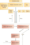
Connections between the lumbosacral spinal cord and brain areas involved in bladder control. The central pathways involved in controlling the urinary bladder can be visualized in rats using transneuronal virus tracing. Injection of pseudorabies virus into the wall of the urinary bladder leads to retrograde transport of the virus (indicated by the dashed arrows) and the sequential infection of postganglionic neurons, preganglionic neurons, spinal interneurons, and then various supraspinal neural circuits that are synaptically linked to the spinal preganglionic neurons and interneurons. The supraspinal sites labeled by the virus transport include the pontine micturition centre (also known as Barrington’s nucleus), the cerebral cortex, the paraventricular nucleus (PVN), the medial preoptic area (MPOA) and periventricular nucleus (PeriVN) of the hypothalamus, the periaqueductal gray (PAG), the locus coeruleus (LC) and subcoeruleus, the red nucleus (Red N.), the raphe nuclei, and the A5 noradrenergic cell group. Synaptic connections are indicated by solid arrows. Synaptic inputs from supraspinal neurons can project to spinal preganglionic neurons or interneurons, as indicated by the bracket (216).
Figure 12
Figure 12
Neural circuits that control continence and micturition. (A) Urine storage reflexes. During the storage of urine, distention of the bladder produces low-level vesical afferent firing. This in turn stimulates the sympathetic outflow in the hypogastric nerve to the bladder outlet (the bladder base and the urethra) and the pudendal outflow to the external urethral sphincter. These responses occur by spinal reflex pathways and represent guarding reflexes, which promote continence. Sympathetic firing also inhibits contraction of the detrusor muscle and modulates neurotransmission in bladder ganglia. A region in the rostral pons (the pontine storage centre) might increase striated urethral sphincter activity. (B) Voiding reflexes. During the elimination of urine, intense bladderafferent firing in the pelvic nerve activates spinobulbospinal reflex pathways (shown in blue) that pass through the pontine micturition centre. This stimulates the parasympathetic outflow to the bladder and to the urethral smooth muscle (shown in green) and inhibits the sympathetic and pudendal outflow to the urethral outlet (shown in red). Ascending afferent input from the spinal cord might pass through relay neurons in the periaqueductal gray (PAG) before reaching the pontine micturition centre. Note that these diagrams do not address the generation of conscious bladder sensations, nor the mechanisms that underlie the switch from storage to voluntary voiding, both of which presumably involve cerebral circuits above the PAG. R represents receptors on afferent nerve terminals (216).
Figure 13
Figure 13
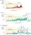
Bladder (top traces) and EUS EMG activity (bottom traces) recorded during a continuous transvesical infusion CMG in an anesthetized rat. (A) Intravesical pressure and EUS EMG activity were relatively stable during the filling phase. A reflex bladder contraction, indicated by an abrupt, large increase in bladder pressure, was accompanied by large-amplitude EUS EMG activity. (B) Same recording indicated by asterisk in A shown at faster time scale. The bracket in B indicates the recording period in C, and the bracket in C indicates the recording period in D at a faster time scale. Note the decline in intravesical pressure during EUS EMG bursting in B and C, which indicates the period of voiding. (C) Tonic EUS EMG activity precedes the large rise in intravesical pressure and shifts to a bursting pattern at the peak of bladder contraction before the onset of voiding. Small oscillations in intravesical pressure coincide with each burst of EMG activity. (D) Recordings in C shown at very fast time scale showing individual EUS EMG bursts composed of active (AP) and silent periods (SP; brackets) and the small fluctuations in intravesical pressure accompanying each burst. Vertical calibration, intravesical pressure (in cmH2O); horizontal calibration, time (in minutes or seconds); Inf, start of saline infusion (104).
Figure 14
Figure 14
Reflex pathways to EUS and bladder in rat in spinal intact (A) and after chronic thoracic spinal cord transection (SCT) (B); (C) pelvic afferent to EUS reflexes. (A) Diagram showing putative reflex pathways mediating reflex micturition and tonic and bursting EUS activity in spinal cord-intact (A) and chronic T8–9 SCT rats (B). (A) Spinobulbospinal micturition reflex pathway is shown by the solid line passing through the pontine micturition center (PMC) in the rostral brain stem. The hypothesized pathway mediating EUS bursting is shown by the dotted line also passing through the PMC. In spinal cord-intact rats, when the bladder is distended, afferent input from bladder mechanoreceptors passes via the pelvic nerve to the L6-S1 spinal cord to the spinal EUS-control center to generate tonic EUS activity and the ER. Input from the L6-S1 spinal cord passes to the PMC, which then projects to the lumbosacral micturition center to generate reflex bladder contractions and to L3-4 bursting center to generate EUS bursting. The spinal EUS bursting center provides an excitatory input to the spinal EUS-control center to initiate an excitatory outflow to the EUS. The spinal EUS-control center in the L6-S1 spinal cord consists of interneuronal and motoneuronal circuitry that regulates EUS activity. (B) After SCT, descending input from the PMC to spinal centers is interrupted. This initially eliminates the micturition reflex, the long latency EUS late reflex (LR), and EUS bursting. The short latency EUS early reflex (ER) and tonic EUS activity mediated by a spinal reflex pathway are preserved. However, in chronic SCT rats it is hypothesized that reorganization of synaptic connections in the spinal cord leads to the reemergence of the micturition reflex as well as the LR and EUS bursting. This reorganization depends on the formation of new pathways between pelvic primary afferent nerves and the L3-4 spinal EUS bursting center (dotted line) and spinal micturition center (solid line) or upregulation of pathways that exist in the spinal intact animals. (C) Pelvic afferent to EUS reflexes in spinal intact (top), acute SCI (middle), and chronic SCI rats (bottom tracing). Early reflex (ER) persists after acute SCI but late reflex (LR) is abolished. However, LR recovers in SCT rats if connections between L3-L4 and L6-S1 spinal segments are intact (98).
Figure 15
Figure 15
(A) A representation (prior to the advent of functional brain imaging) of cerebral areas involved in micturition. + = facilitation, − = inhibition; ac = anterior cingulate gyrus, am = amygdala, pl = paracentral lobule, po = preoptic nucleus, rf = pontine reticular formation, sc = subcallosal cingulate gyrus, se = septal area, sfg = superior frontal gyrus. (B) Sagittal section (8 mm off midline) showing medial frontal region (yellow) and presumptive PMC (small yellow region in brainstem) activated during voiding (70, 647).
Figure 16
Figure 16
(A) Brainstem and midbrain areas activated during withholding of urine or with full bladder, or during voiding, projected on a lateral view of the brain. Based on PET, fMRI, and SPECT studies in healthy controls, adapted from Ref. (with permission). (B) In urgency-incontinent women, regions activated (a, yellow) and deactivated (b, blue) during the sensation of urgency. SMA = supplementary motor area; SFG = superior frontal gyrus; dACG = dorsal anterior cingulate gyrus; RI = right insula; dlPFC = dorsolateral prefrontal cortex; PFC = prefrontal cortex (ventromedial or medial) [A (217,241), B (609)).
Figure 17
Figure 17
Coronal (A), sagittal (B), and axial (C) location of lesions causing incontinence (or occasionally retention) in the group of patients studied by Andrew and Nathan (21). The red ellipse shows where white-matter lesions caused lasting urinary tract dysfunction. The cyan ellipse shows the location of gray-matter lesions that caused transient dysfunction [Nathan, personal communication with Dr. Clare Fowler (217)].
Figure 18
Figure 18
Top and left panels (red/yellow): subcortical and temporal regions active in normal controls at low bladder volume when there is little bladder sensation, possibly representing circuit 3 in Figure 8; bottom right panel (blue): similar regions where activity changes after improvement of sensation by sacral neuromodulation, in women with Fowler’s syndrome. (Blue image, 308; orange image, our own unpublished work.)
Figure 19
Figure 19
Multiunit recordings of reflex activity on a bladder postganglionic nerve in a chloralose anesthetized cat during electrical stimulation (0.8 Hz, 3 v, 0.05 ms duration) of bladder afferent axons in the pelvic nerve. The bladder was distended with saline to a volume below the threshold for inducing micturition. First tracing in the upper right is a recording prior to the onset of stimulation showing that the efferent pathway is inactive. The next tracing shows lack of a response to the first stimulus in a train of stimuli. Further stimulation (lower tracings) induces a gradual increase in the magnitude of a long latency reflex and the eventual emergence of asynchronous firing (last tracing) which indicates the onset of reflex micturition. The diagram on the left shows the spinobulbospinal micturition reflex pathway and the sites of nerve stimulation and recording (173).
Figure 20
Figure 20
Relationship between single unit activity in the PMC of a decerebrate, unanesthetized cat, and reflex contractions of the urinary bladder. Top tracings are blood pressure, middle tracings are ratemeter recordings of unit activity in spikes per second and the bottom tracings are bladder pressure in cm H2O. Three types of neuronal activity are illustrated: (A) a direct neuron that only fired during a bladder contraction, (B) an inverse neuron that fired between bladder contractions and was inhibited during contractions, and (C) an independent neuron that exhibited continuous firing unrelated to bladder contractions. Small increases in blood pressure occurred during bladder contractions. The bladder was distended with saline and maintained under isovolumetric conditions. Horizontal calibration represents 1 min. The three neurons were studied at different times in the same animal (173).
Figure 21
Figure 21
Blood oxygen level-dependent (BOLD) images showing brain stem activation associated with switching from the bladder storage phase to the bladder contraction phase. The locations of coronal brain sections (F–G) are indicated in the sagittal brain image at the bottom, which correspond to the Bregma coordinates in the anterior-posterior direction at 2.28, 0.24, 1.80, 3.84, 5.88, 7.80, and 9.84 mm. Region of interest (ROI) analysis was performed on the brain stem at coronal sections F and G to detect the activation. The periaqueductal gray (PAG) and pontine micturition center (PMC) are indicated by the blue arrows. The color scale bars indicate the t value (612).
Figure 22
Figure 22
Computer model of PMC-PAG switching circuits. Diagram illustrating the putative pathways in the periaqueductal gray (PAG) and pontine micturition center (PMC) that contribute to urine storage and voiding. This circuitry shows the neuronal elements and connections used in the computer model. The right side illustrates the ascending afferent limb of the spinobulbospinal micturition reflex that projects to the PAG, and the left side shows the descending limb that connects the PMC direct neuron to the bladder efferent neuron in the sacral spinal cord. During urine storage as the bladder slowly fills low level of afferent activity activates an excitatory neuron (E) in the PAG which relays information (pathway A) to an inverse neuron (I) in the PMC that in turn provides inhibitory input to the type 1 direct neuron (D) to maintain continence. Bladder afferent input is also received by a second neuron in the PAG (E) that is on the excitatory pathway (pathway B) to the PMC type 1 direct neuron (D) and to a transiently active PMC neuron (T) that fires at the beginning of micturition. However, the PAG excitatory relay neuron (E) is not activated during the early stages of bladder filling because it is inhibited by a tonically active independent neuron (I). The PMC type 1 direct neuron is also inhibited by a tonically active independent neuron (I) located in the PMC. Bladder afferent firing gradually increases during bladder filling which increases feedforward inhibition of the direct neuron via the PAG-PMC inverse neuron pathway. However, at a critical level of afferent firing, excitatory input to the PAG excitatory relay neuron surpasses the tonic inhibition of the independent neuron and sends signals to the PMC transient neuron which briefly inhibits the inverse neuron reducing inhibitory input to the direct neuron allowing it to overcome tonic inhibition and fire action potentials which activate by an axon collateral (pathway C) a reciprocal inhibitory neuron (R) that suppresses the inverse neuron (I) and further reduces inhibition of the direct neuron (D). The direct neuron then switches into maximal firing mode and sends excitatory input to the spinal efferent pathway to the bladder inducing a large bladder contraction and more afferent firing which further enhances synaptic transmission in the PAG-PMC micturition reflex pathways. The reflex circuitry returns to storage mode as the bladder empties and afferent firing declines. Excitatory neurons are green and inhibitory neurons are red (173).
Figure 23
Figure 23
Computer simulation of the storage-voiding cycle. Simulated bladder volume (top tracing) and pressure (second tracing), bladder afferent firing (third tracing) and bladder efferent firing (bottom tracing) during bladder filling (30 mL/min) and during reflex voiding using the computer model of spinal, PAG and PMC neural pathways and the myocybernetic model of Bastiaanssen et al. 1996 to predict the properties of the bladder, urethra, and the afferent firing arising in these structures. Note that as bladder volume increases, bladder pressure remains low, bladder efferent firing is absent, but bladder afferent firing gradually increases eventually reaching a threshold for inducing a micturition reflex as evidenced by an abrupt increase in efferent firing, which induces an increase in bladder pressure, increased afferent firing and bladder emptying. Bladder efferent firing peaks early during micturition and is maintained until the bladder is empty. The voiding phase is shown on an expanded time scale in the tracings on the right side (173).
Figure 24
Figure 24
Hypothetical diagram showing dopaminergic and adenosinergic mechanisms inducing bladder dysfunction in Parkinson’s disease (PD). The micturition reflex is controlled by the spinobulbospinal pathway passing through the PAG in the midbrain and the PMC in the pons. This neural circuit is under the control of higher centers including the striatum and the cortex region. A. Under normal conditions (Intact), tonic firing (+) of dopaminergic neurons in the SN activates dopamine D1 receptors expressed on GABAergic inhibitory neurons in the striatum to induce tonic GABAergic (−) inhibition of the micturition reflex at the level of the PAG. At the same time, D1 receptor stimulation suppresses the activity of adenosinergic neurons, which exert an excitatory effect on micturition via adenosine A2A receptors [Adenosine A2A (+)]. B. In PD, dopaminergic neurons in the SN are lost (lesion), leading to the loss of dopamine D1 receptor activation [D1 (loss of activation)], which results in reduced activation of inhibitory GABAergic neurons in the striatum [GABA (loss of inhibition)]. At the same time, reduced D1 receptor stimulation enhances the adenosinergic mechanism to stimulate adenosine A2A receptors [Adenosine A2A (++)], leading to facilitation of the spinobulbospinal pathway controlling the micturition reflex (activity). Administration of dopamine D1 receptor agonists (D1 agonist) can restore the GABAergic nerve activity and suppress A2A receptor-mediated activation to reduce bladder overactivity in PD. Also, administration of adenosine A2A receptor antagonists (A2A antagonist) can suppress A2A receptor-mediated activation of the micturition reflex to reduce bladder overactivity in PD. Dopamine D2 receptors [D2 (+)] expressed in the spinal cord enhance the micturition reflex. Abbreviations: dopamine D1 receptor (D1); dopamine D2 receptor (D2); gamma-aminobutyric acid (GABA); periaqueductal gray (PAG); pontine micturition center (PMC); substantia nigra pars compacta (SN).
Figure 25
Figure 25
Organization of the parasympathetic excitatory reflex pathway to the detrusor muscle. This scheme is based on results from electrophysiological studies in cats. Micturition is initiated by a supraspinal reflex pathway that passes through a center in the brainstem. The pathway is triggered by myelinated afferents (Aδ-fibers), which are connected to the tension receptors in the bladder wall. Injury to the spinal cord above the sacral segments interrupts the connections between the brain and spinal autonomic centers and initially blocks micturition. However, following cord injury a spinal reflex mechanism (shown in green) emerges that is triggered by unmyelinated vesical afferents (C-fibers); the A-fiber afferent inputs are ineffective. The C-fiber reflex pathway is usually weak or undetectable in animals with an intact nervous system. Stimulation of the C-fiber bladder afferents by instillation of ice water into the bladder (cold stimulation) activates voiding responses in patients with spinal cord injury. Capsaicin (20-30 mg, subcutaneously) blocks the C-fiber reflex in cats with spinal lesions but does not block micturition reflexes in spinal intact cats. Intravesical capsaicin also suppresses detrusor hyperreflexia and cold-evoked reflexes in patients with neurogenic bladder dysfunction (216).
Figure 26
Figure 26
Peripheral mechanisms involved in the neurotrophin-mediated development of bladder overactivity. In urinary bladder, NGF (shown in blue) is produced by several cell types—including urothelium, mast cells, and detrusor smooth muscle cells—upon stretch or inflammation. The urothelium also potentially produces BDNF (shown in red). NGF binding to TrkA receptors on the urothelium might directly activate urothelial sensory ion channels, such as TRPV1 (shown in purple), or increase expression of TRPV1 and mechanosensitive channels (MSC, shown in pink). Increased TRPV1 and MSC activity stimulate the release of urothelial mediators, such as ATP, which sensitize the underlying afferents. In addition, NGF activates TrkA receptors expressed on suburothelial afferent C-fiber terminals, directly sensitizing neuronal TRPV1, MSCs and voltage-gated ion channels (VGCs, shown in orange). The TrkA-NGF complex is internalized (dashed lines) and retrogradely transported to cell bodies in lumbosacral DRG, where de novo transcription of TRPV1, VGCs, MSCs and additional sensory ion channels (including purinergic P2X3 receptor for ATP; shown in green) is initiated. These newly synthesized ion channels are anterogradely transported back to afferent terminals to contribute to peripheral hypersensitivity. Neurotrophin receptors TrkB (shown in red) and p75NTR (shown in black) are also expressed on both urothelium and afferent terminals, although their role has not yet been defined. Abbreviations: ATP, adenosine triphosphate; BDNF, brain-derived nerve factor; BOO, bladder outlet obstruction; BPS, bladder pain syndrome; DRG, dorsal root ganglia; MSC, mechanosensory channel; NGF, nerve growth factor; OAB, overactive bladder syndrome; P2X3, P2X purinoceptor 3; TrkA, tropomyosin-related kinase A; TrkB, tropomyosin-related kinase B; TRPV1, transient receptor potential cation channel vanilloid subfamily member 1; VGC, voltage-gated ion channel (486).
Figure 27
Figure 27
Central mechanisms involved in the neurotrophin-mediated development of bladder overactivity. Upon retrograde transport of TrkA-NGF complexes along the afferent fibers from the bladder, DRG neurons increase synthesis of excitatory neuromediators, such as substance P, CGRP, BDNF, and voltage-gated ion channels, which are transported anterogradely to primary afferent terminals in the lumbosacral spinal cord. Following the synaptic release, substance P (shown in green), CGRP (shown in brown), and BDNF (shown in red) activate their corresponding receptors (NK1, CGRP receptor, and TrkB, respectively) to induce central sensitization of nociceptive, and possibly also micturition, pathways. One of the mechanisms of central sensitization involves BDNF-induced activation of the NMDA receptor for excitatory mediator glutamate (shown in gray). Enhanced voltage-gated ion channel activity could contribute towards increased firing of bladder afferents. NGF and TrkA are also detected in the spinal cord, but their origin and function remain unknown. Following sensitization and activation of the central (spinobulbospinal) micturition reflex, excessive efferent stimulation could eventually contribute to DO. Abbreviations: BDNF, brain-derived nerve factor; CGRP, calcitonin gene-related peptide; CGRPR, calcitonin gene-related peptide receptor; DO, detrusor overactivity; DRG, dorsal root ganglia; Glu, Glutamate; NGF, nerve growth factor; NK1, neurokinin-1 receptor; NMDA, N-methyl-D-aspartate; MPG, major pelvic ganglia; SP, substance P; Trk, tropomyosin-related kinase; VGC, voltage-gated ion channel (486).

References

    1. Abdel-Gawad M, Dion SB, Elhilali MM. Evidence of a peripheral role of neurokinins in detrusor hyperreflexia: A further study of selective tachykinin antagonists in chronic spinal injured rats. J Urol. 2001;165:1739–1744. - PubMed
    1. Abdel-Karim AM, Barthlow HG, Bialecki RA, Elhilali MM. Effects of M274773, a neurokinin-2 receptor antagonist, on bladder function in chronically spinalized rats. J Urol. 2005;174:1488–1492. - PubMed
    1. Abe F. Inhibitory presynaptic effect of noradrenaline on the hypogastric ganglion of the guinea pig. J Pharmacol Exp Ther. 1984;231:395–403. - PubMed
    1. Abrams P, Cardozo L, Fall M, Griffiths D, Rosier P, Ulmsten U, van Kerrebroeck P, Victor A, Wein A, Standardisation Sub-committee of the International Continence S The standardisation of terminology of lower urinary tract function: Report from the Standardisation Subcommittee of the International Continence Society. Neurourol Urodyn. 2002;21:167–178. - PubMed
    1. Adams PR, Brown DA, Constanti A. M-currents and other potassium currents in bullfrog sympathetic neurons. J Physiol. 1982;330:537–572. - PMC - PubMed

MeSH terms

Substances