. 2015 Jan 15:5:7798.
doi: 10.1038/srep07798.

The CarO rhodopsin of the fungus Fusarium fujikuroi is a light-driven proton pump that retards spore germination

Affiliations

The CarO rhodopsin of the fungus Fusarium fujikuroi is a light-driven proton pump that retards spore germination

Jorge García-Martínez et al. Sci Rep. .

Abstract

Rhodopsins are membrane-embedded photoreceptors found in all major taxonomic kingdoms using retinal as their chromophore. They play well-known functions in different biological systems, but their roles in fungi remain unknown. The filamentous fungus Fusarium fujikuroi contains two putative rhodopsins, CarO and OpsA. The gene carO is light-regulated, and the predicted polypeptide contains all conserved residues required for proton pumping. We aimed to elucidate the expression and cellular location of the fungal rhodopsin CarO, its presumed proton-pumping activity and the possible effect of such function on F. fujikuroi growth. In electrophysiology experiments we confirmed that CarO is a green-light driven proton pump. Visualization of fluorescent CarO-YFP expressed in F. fujikuroi under control of its native promoter revealed higher accumulation in spores (conidia) produced by light-exposed mycelia. Germination analyses of conidia from carO(-) mutant and carO(+) control strains showed a faster development of light-exposed carO(-) germlings. In conclusion, CarO is an active proton pump, abundant in light-formed conidia, whose activity slows down early hyphal development under light. Interestingly, CarO-related rhodopsins are typically found in plant-associated fungi, where green light dominates the phyllosphere. Our data provide the first reliable clue on a possible biological role of a fungal rhodopsin.


Figures

Figure 1
Figure 1. Schematic representation of CarO::YFP.
Topo2 transmembrane protein display software and NCBI protein-Blast was used to define transmembrane sections in CarO (GenBank: CAD97459.1) in analogy to BR (SWISS Model, template 4hwl.3.B). Conserved residues are highlighted in orange and related residues in yellow. Asp117 and Asp128 in helix-C, highlighted in red, represent the putative proton acceptor and donor, respectively (D85 and D96 in BR). The lysine (Lys246) in helix-G is also conserved and allows for covalent binding of retinal via protonated Schiff base. In helix-D the Trp148 (red font) is typical for auxiliary ORP-like rhodopsins, which contain Glu or Trp in this positions instead of Gly in BR and other related rhodopsins. A linker peptide (gray letters) was used to connect YFP to the C-terminus of the CarO protein.
Figure 2
Figure 2. Electrophysiological analysis of CarO::YFP expressed in Flip-In T-Rex 293 cells.
(a) Wide field fluorescence image of Flip-In T-Rex 293 CarO::YFP cells used in patch-clamp experiments. White bar represents 10 μm. The fusion protein is mainly located in the plasma membrane but also in internal membranes. (b) Typical current traces of the ion pump CarO recorded in NaCl pH 7.4 at membrane potentials of +40 mV, 0 mV, and −100 mV, respectively, as indicated. Cells were illuminated by a 561 nm diode pumped laser. The green bar represents illumination time. (c) I–V-relationship of the light-dependent normalized pump current of CarO::YFP measured at pH 7.4 corresponding to data shown in b. Note, that activity of CarO is voltage-dependent.
Figure 3
Figure 3. Light dependency of CarO::YFP.
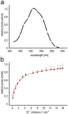
(a) Action spectrum. Effect of light-wavelength on CarO::YFP activity (mean value and standard deviation of 7 cells). The light emitted by a 150W-XBO lamp was filtered through narrow-band filters. At every wavelength the same dose of photons per time and area (2 × 10¹⁶ photons s−1 mm−2) was used and values were corrected for eventual intensity loss (reference wavelength 560 nm). The action spectrum was essentially the same at different intracellular proton concentrations (pH 5, 7.4, and 9, Supplementary Fig. S2); therefore, for better signal to noise-ratio, pH of internal pipette solution was set to 5. (b) Dependency of the pump current amplitude on photon density. Mean normalized photocurrent and corresponding standard deviation (5 cells, 3 recordings each). Data were fitted by a Hill function (red curve).
Figure 4
Figure 4. Pump-activity of CarO::YFP at various pHs and other ion concentrations.
Cells were illuminated by a 561 nm-laser. (a) Influence of external pH on pump-activity. The relative pump activity is shown, which was normalized to the pump current at pH 7.4 and 0 mV. pH of bath solution varied from pH 5 (black squares, 10 cells), to pH 7.4 (red circles, 25 cells) and pH 9 (blue triangles, 13 cells); intracellular pH was kept at 7.4. (b) Influence of internal pH on pump activity. Extracellular pH was maintained at pH 7.4. Mean area specific photocurrent at intracellular pH 9 (3 cells), 7.4 (8 cells), and 5 (10 cells), respectively. (c) Analysis of pump activity in absence of extracellular chloride or intracellular sodium. Mean area-specific pump current to membrane voltage relationship of CarO is given. Sodium chloride in standard solutions (red circles, 14 cells) was replaced either intracellularly by potassium chloride (black squares, 5 cells) or extracellularly by sodium sulfate (blue triangles, 7 cells), without any remarkable effect on pump activity.
Figure 5
Figure 5. Influence of weak organic acids on the pump-activity of CarO::YFP.
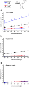
I–V blot of the normalized light-driven pump current (linear fit of mean values and standard deviation of measurements recorded from at least 5 different cells) at pH 5 (black squares), pH 7.4 (red circles) and pH 9 (blue triangles), respectively, in extracellular presence of the weak organic acids gluconate (a), glutamate (b), and galacturonate (c). Note that in the presence of gluconate and glutamate at pH 5 the pump activity is enhanced, though the contra-directed gradient is increased. In case of gluconate at pH 9 a transient enhanced pump current (half triangles, pH 9*) is observed, which disappears in a time scale of over ten minutes (closed triangles).
Figure 6
Figure 6. Localization of CarO::YFP in F. fujikuroi.
(a) Southern blot analysis of genomic DNA of F. fujikuroi FKMC1995 transformed with pHJA2-carO::YFP and selected for hygromycin resistance. DNA samples were digested with NcoI or BclI. The carO::yfp gene was introduced into the fungal genome and the native carO gene was not replaced. Complete blot is shown in the Supplementary Fig. S13. (b) Localization of CarO::YFP in F. fujikuroi conidia. The CarO::YFP fusion protein is mainly located in the plasma membrane but also in the membranes of inner organelles (vacuole, nucleus, ER). i–iii. Photomicrographs of conidia obtained by transmitted light microscopy (upper row) and corresponding confocal laser scanning microscopy (cLSM; lower row). The expression of carO::yfp in the conidia is induced by light. i) Light exposed (left: bicellular, right, monocellular), ii) dark exposed, iii) control (light exposed wild type). White bars represent 5 μm (i) and 2 μm (ii, iii), respectively. (c) Confocal laser scanning microphotograph of a yeast cell (S. cerevisiae DSY5) expressing CarO::YFP. The fusion protein is mainly located in the plasma membrane but in addition also in internal membranes (most likely ER). White bar represents 1 μm. (d) Localization of CarO::YFP in light-exposed F. fujikuroi hyphae 20 h after germination. The CarO::YFP fusion protein is heterogeneously distributed in the mycelia according to a dynamic process (Supplementary Fig. S6 and Movie 1). White bar represents 5 μm.
Figure 7
Figure 7. Colony growth of F. fujikuroi carO and carO+ strains in the dark and under light.
(a) carO+ and carO strains were spotted in standard DA minimal medium (104 conidia per drop) and documented after 2 days incubation under light. White bar represents 5 mm. (b) Influence of carbon source on colony size. Inoculation was done as described in (a) but in DA media with varying additional carbon sources as indicated. Colonies were either grown in the light or in the dark. (c) Linear growth experiments in race tubes. carO+ and carO strains were grown in standard DA minimal medium (104 conidia per drop; n = 4) and colony length was measured over a period of 9 days. Mean values are given with standard deviation (y-bars).
Figure 8
Figure 8. Influence of CarO on conidia germination.
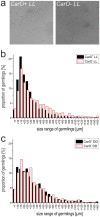
Conidia from light and dark exposed mycelia of the CarO+ and CarO strains were seeded in liquid DA medium adjusted to pH 5 and germination was observed. Data from one single representative experiment are shown, but similar results were obtained in four independent experiments. (a) Representative images of germinated conidia of light exposed strains. (b) Length of germlings of CarO+ LL (n = 1476) and CarO LL (n = 1413) conidia as indicated. The percentage of >40 μm germlings is higher in CarO LL than in CarO+ LL, while the opposite is the case for <30 μm germlings. (c) Length of germlings of CarO+ DD (n = 1392) and CarO DD (n = 1196) conidia as indicated. Only slight difference was observed between both strains incubated in the dark.

References

    1. Corrochano L. M. Fungal photoreceptors: sensory molecules for fungal development and behaviour. Photochem. Photobiol. Sci. 6, 725–736 (2007). - PubMed
    1. Bayram Ö., Braus G. H., Fischer R. & Rodríguez-Romero J. Spotlight on Aspergillus nidulans photosensory systems. Fungal Genet. Biol. 47, 900–908 (2010). - PubMed
    1. Idnurm A., Verma S. & Corrochano L. M. A glimpse into the basis of vision in the kingdom Mycota. Fungal Genet. Biol. 47, 881–892 (2010). - PMC - PubMed
    1. Avalos J. & Estrada A. F. Regulation by light in Fusarium. Fungal Genet. Biol. 47, 930–938 (2010). - PubMed
    1. Herrera-Estrella A. & Horwitz B. A. Looking through the eyes of fungi: molecular genetics of photoreception. Mol. Microbiol. 64, 5–15 (2007). - PubMed

Publication types

MeSH terms

LinkOut - more resources