

The tissue distribution of lipoprotein lipase determines where chylomicrons bind
- PMID: 25589507
- PMCID: PMC4340306
- DOI: 10.1194/jlr.M056028
The tissue distribution of lipoprotein lipase determines where chylomicrons bind
Abstract
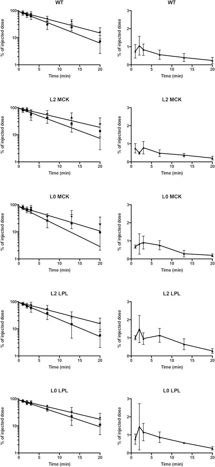
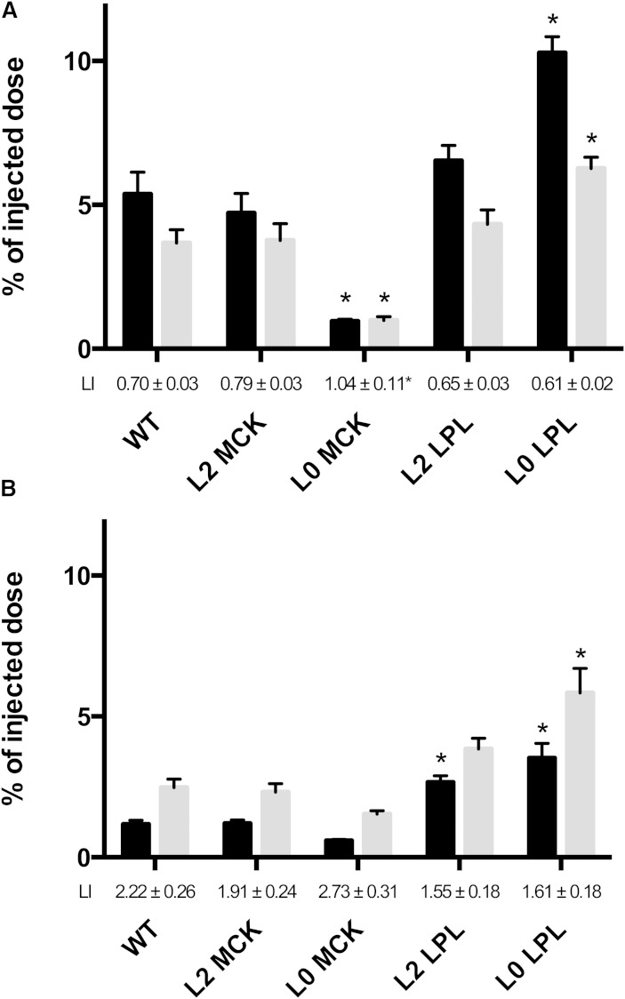
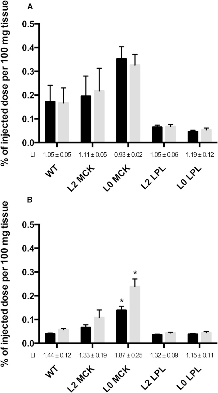
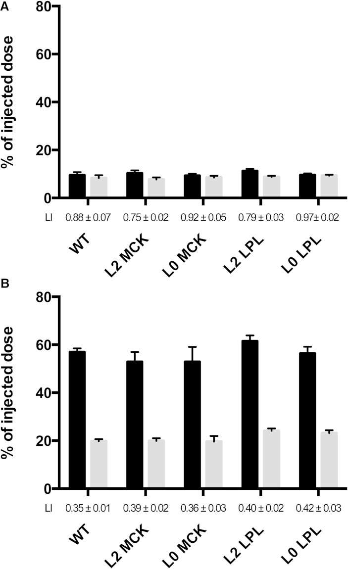
To determine the role of LPL for binding of lipoproteins to the vascular endothelium, and for the distribution of lipids from lipoproteins, four lines of induced mutant mice were used. Rat chylomicrons labeled in vivo with [(14)C]oleic acid (primarily in TGs, providing a tracer for lipolysis) and [(3)H]retinol (primarily in ester form, providing a tracer for the core lipids) were injected. TG label was cleared more rapidly than core label. There were no differences between the mouse lines in the rate at which core label was cleared. Two minutes after injection, about 5% of the core label, and hence chylomicron particles, were in the heart of WT mice. In mice that expressed LPL only in skeletal muscle, and had much reduced levels of LPL in the heart, binding of chylomicrons was reduced to 1%, whereas in mice that expressed LPL only in the heart, the binding was increased to over 10%. The same patterns of distribution were evident at 20 min when most of the label had been cleared. Thus, the amount of LPL expressed in muscle and heart governed both the binding of chylomicron particles and the assimilation of chylomicron lipids in the tissue.
Keywords: adipose tissue; heart; liver; muscle; nonesterified fatty acids; transgene; triglycerides.
Copyright © 2015 by the American Society for Biochemistry and Molecular Biology, Inc.
Figures
References
-
- Wang H., Eckel R. H. 2009. Lipoprotein lipase: from gene to obesity. Am. J. Physiol. Endocrinol. Metab. 297: E271–E288. - PubMed
-
- Olivecrona T., Olivecrona G. 2009. The ins and outs of adipose tissue. In Cellular Lipid Metabolism. C. Ehnholm, editor. Springer, New York. 315–369.
-
- Hultin M., Savonen R., Olivecrona T. 1996. Chylomicron metabolism in rats: lipolysis, recirculation of triglyceride-derived fatty acids in plasma FFA, and fate of core lipids as analyzed by compartmental modelling. J. Lipid Res. 37: 1022–1036. - PubMed
-
- Cooper A. D. 1997. Hepatic uptake of chylomicron remnants. J. Lipid Res. 38: 2173–2192. - PubMed
Publication types
MeSH terms
Substances
LinkOut - more resources
Full Text Sources
Other Literature Sources
Molecular Biology Databases
Miscellaneous
