. 2015 Mar;89(6):3366-79.
doi: 10.1128/JVI.03111-14. Epub 2015 Jan 14.

Absence of the uracil DNA glycosylase of murine gammaherpesvirus 68 impairs replication and delays the establishment of latency in vivo

Affiliations

Absence of the uracil DNA glycosylase of murine gammaherpesvirus 68 impairs replication and delays the establishment of latency in vivo

Nana Minkah et al. J Virol. 2015 Mar.

Abstract

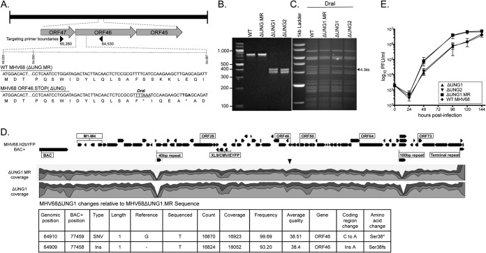
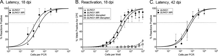
Uracil DNA glycosylases (UNG) are highly conserved proteins that preserve DNA fidelity by catalyzing the removal of mutagenic uracils. All herpesviruses encode a viral UNG (vUNG), and yet the role of the vUNG in a pathogenic course of gammaherpesvirus infection is not known. First, we demonstrated that the vUNG of murine gammaherpesvirus 68 (MHV68) retains the enzymatic function of host UNG in an in vitro class switch recombination assay. Next, we generated a recombinant MHV68 with a stop codon in ORF46/UNG (ΔUNG) that led to loss of UNG activity in infected cells and a replication defect in primary fibroblasts. Acute replication of MHV68ΔUNG in the lungs of infected mice was reduced 100-fold and was accompanied by a substantial delay in the establishment of splenic latency. Latency was largely, yet not fully, restored by an increase in virus inoculum or by altering the route of infection. MHV68 reactivation from latent splenocytes was not altered in the absence of the vUNG. A survey of host UNG activity in cells and tissues targeted by MHV68 indicated that the lung tissue has a lower level of enzymatic UNG activity than the spleen. Taken together, these results indicate that the vUNG plays a critical role in the replication of MHV68 in tissues with limited host UNG activity and this vUNG-dependent expansion, in turn, influences the kinetics of latency establishment in distal reservoirs.

Importance: Herpesviruses establish chronic lifelong infections using a strategy of replicative expansion, dissemination to latent reservoirs, and subsequent reactivation for transmission and spread. We examined the role of the viral uracil DNA glycosylase, a protein conserved among all herpesviruses, in replication and latency of murine gammaherpesvirus 68. We report that the viral UNG of this murine pathogen retains catalytic activity and influences replication in culture. The viral UNG was impaired for productive replication in the lung. This defect in expansion at the initial site of acute replication was associated with a substantial delay of latency establishment in the spleen. The levels of host UNG were substantially lower in the lung compared to the spleen, suggesting that herpesviruses encode a viral UNG to compensate for reduced host enzyme levels in some cell types and tissues. These data suggest that intervention at the site of initial replicative expansion can delay the establishment of latency, a hallmark of chronic herpesvirus infection.


Figures

FIG 1
FIG 1
MHV68 UNG complements UNG−/− B lymphocytes for class switch recombination (CSR). (A) Representative flow cytometry plot demonstrating that WT, but not UNG−/− B cells undergo CSR to IgG1. (B) Schematic of CSR assay. Primary B cells from the indicated naive mice are treated with LPS and IL-4 for 4 days. (C) Representative flow cytometry plot of CSR to IgG1 in UNG−/− splenocytes transduced with empty vector or pMX expressing murine UNG2, MHV68 vUNG, or MHV68 vUNG.stop. The percentage of infected (GFP+) cells expressing IgG1 is indicated on the plot. (D) Summary graph of percentage of GFP+ cells that underwent CSR to IgG1 4 days after retroviral infection. Bars represent the means ± the standard deviations (*, P ≤ 0.05; **, P ≤ 0.005). UNG complementation data are representative of three independent experiments.
FIG 2
FIG 2
Construction and characterization of a recombinant MHV68ΔUNG virus. (A) Schematic of the ΔUNG virus. WT and mutant ORF46 nucleotide sequences are compared, highlighting the presence of the unique DraI site and insertion of the TAA stop codon in ΔUNG (underlined). Underneath the nucleotide sequence is the translated amino acid sequence. An asterisk (*) indicates the presence of the stop codon. Arrows indicate the boundaries of primers to generate a 725-bp amplimer to verify the mutation. (B) DraI digestion of a 725-bp amplimer to confirm the unique DraI site in the two independent ORF46.STOP viruses (ΔUNG1 and ΔUNG2), but not the marker rescue of ΔUNG1 (ΔUNG1.MR) or wild-type (WT) MHV68. (C) Confirmation of mutant and repaired MHV68 viruses by restriction fragment length polymorphism analysis. Portions (10 μg) of WT BAC DNA, ΔUNG1.MR, ΔUNG1, or ΔUNG2 were digested with DraI and resolved on a 0.8% agarose gel. An arrowhead indicates the loss of a 4.3-kb fragment with the insertion of the unique DraI site into ORF46. (D) Analysis of MHV68 BAC by whole-genome sequencing. (Top) Illumina reads were aligned to the MHV68.H2bYFP BAC genome. Shown in the histogram are levels of coverage across the genome; the scale is 0 to ×30,000, 56 million total mapped reads for ΔUNG1, and 39 million reads for ΔUNG1.MR. Variants are indicated by inverted triangles. The two regions of low coverage are due to the paucity of uniquely mapped reads in the small repeat regions. (Bottom) Table of expected variants in the UNG.STOP BAC not found in UNG.MR. (E) Multistep growth curve in primary MEF cells at an MOI of 0.01 with the indicated viruses.
FIG 3
FIG 3
The vUNG of MHV68 promotes replication in primary fibroblasts. (A) Single-step growth curve in immortalized NIH 3T3 fibroblasts at an MOI of 5.0 with ΔUNG1 and ΔUNG1.MR. (B) Single-step growth curve in primary MEFs at an MOI of 5.0 with ΔUNG1 and ΔUNG1.MR. (C) Relative increase in viral DNA levels from ΔUNG1- or ΔUNG1.MR-infected MEFs at 24 hpi at an MOI of 5. Data are normalized to the 6 hpi input DNA. (D) Time course analysis of early (ORF59) and late (ORF65 and ORF75C) gene products upon a high MOI infection of MEFs. (E) Immunoblot analysis of ORF75C tegument protein levels delivered with incoming virus 1 hpi. The increase in GAPDH-normalized ORF75C levels relative to the MR virus is indicated below the gel.
FIG 4
FIG 4
The vUNG of MHV68 retains enzymatic UNG activity in infected primary fibroblasts. (A) Schematic of UNGase assay using an Alexa 488-labeled oligonucleotide containing a single uracil. Uracil excision leads to oligonucleotide cleavage. (B) Denaturing polyacrylamide gel analysis of oligonucleotide cleavage upon incubation with lysates prepared from MEFs uninfected or infected with WT, ΔUNG1.MR, and ΔUNG1 at 24 hpi. (C) Time course analysis of UNG activity within primary MEFs infected with WT or ΔUNG1 viruses. The percentage of cleavage relative to the negative control is indicated below each gel.
FIG 5
FIG 5
The vUNG of MHV68 is critical for replication in the lungs of infected mice. C57BL/6 mice were infected at 100 PFU by the intranasal route with two independent viruses encoding stop codon disruptions in ORF46 (ΔUNG1 and ΔUNG2) and the marker rescue virus of ΔUNG1 (ΔUNG1.MR). The titers of lung homogenates from mice were determined by plaque assay; the line indicates the geometric mean titer. Each symbol represents an individual mouse. The dashed line depicts the limit of detection at 50 PFU/ml of lung homogenate (log10 of 1.7). *, P ≤ 0.05; **, P ≤ 0.005.
FIG 6
FIG 6
MHV68 vUNG is essential for the establishment of latency in the spleen at early, but not late times during chronic infection after low-dose intranasal inoculation. C57BL/6 mice were infected at 100 PFU by the intranasal route with the indicated viruses. (A) Weights of spleens harvested 16 dpi (***, P ≤ 0.001). (B) Frequency of splenocytes harboring latent genomes at 16 dpi. (C) Frequency of splenocytes undergoing reactivation from latency upon explant. (D) Frequency of splenocytes harboring latent genomes at 6 weeks postinfection. For the limiting-dilution analyses, curve fit lines were determined by nonlinear regression analysis. Using Poisson distribution analysis, the intersection of the nonlinear regression curves with the dashed line at 63.2% was used to determine the frequency of cells that were either positive for the viral genome or reactivating virus. The data were generated from at least three independent experiments for 16 dpi and from two independent experiments for 42 dpi.
FIG 7
FIG 7
Infection with a higher dose partially overcomes the loss of vUNG. C57BL/6 mice were infected at 100,000 PFU by the intranasal route with the indicated viruses. (A) The titers of lung homogenates from mice were determined by plaque assay; the line indicates the geometric mean titer. Each symbol represents an individual mouse. The dashed line depicts the limit of detection at 50 PFU/ml of lung homogenate or a log10 of 1.7. *, P ≤ 0.05; **, P ≤ 0.005. (B) Weights of spleens harvested from the indicated infections 16 dpi. Each symbol represents an individual mouse. *, P ≤ 0.05; **, P ≤ 0.005. (C) Frequency of splenocytes harboring latent genomes. (D) Frequency of splenocytes undergoing reactivation from latency upon explant. For the limiting-dilution analyses, curve fit lines were determined by nonlinear regression analysis. Using Poisson distribution analysis, the intersection of the nonlinear regression curves with the dashed line at 63.2% was used to determine the frequency of cells that were either positive for the viral genome or reactivating virus. The data were generated from at least three independent experiments.
FIG 8
FIG 8
ORF46 is not essential for the establishment of latency upon direct intraperitoneal inoculation. C57BL/6 mice were infected at 100 PFU by the intraperitoneal route with the indicated viruses. (A) The titers of lung homogenates from mice were determined by plaque assay; the line indicates the geometric mean titer. Each symbol represents an individual mouse. The dashed line depicts the limit of detection at 50 PFU/ml of lung homogenate (log10 of 1.7). *, P ≤ 0.05; **, P ≤ 0.005. (B) Weights of spleens harvested from the indicated infections at 18 dpi. Each symbol represents an individual mouse. (C) Frequency of splenocytes harboring latent genomes at 18 dpi. (D) Frequency of splenocytes spontaneously reactivating from latency at 18 dpi. (E) Proportion of CD95hi of CD19+ B cells in response to the indicated infection. (F) Proportion of CD95hi cells in the virus-infected YFP+ CD19+ B cell subset. *, P ≤ 0.05. (G) Weights of spleens harvested at 42 dpi. (H) Frequency of splenocytes harboring latent genomes at 42 dpi. For the limiting-dilution analyses, curve fit lines were determined by nonlinear regression analysis. The dashed lines represent 63.2%. Using Poisson distribution analysis, the intersection of the nonlinear regression curves with the dashed line was used to determine the frequency of cells that were either positive for the viral genome or reactivating virus. The data were generated from at least three independent experiments for 18 dpi and two independent experiments for 42 dpi.
FIG 9
FIG 9
MHV68 vUNG is not required for latency in the peritoneal exudate compartment after intraperitoneal inoculation. C57BL/6 mice were infected at 100 PFU by the intraperitoneal route with ΔUNG1.MR and ΔUNG1 MHV68 viruses. (A) Frequency of peritoneal exudate cells harboring latent genomes at 18 dpi. (B) Frequency of splenocytes spontaneously reactivating from latency at 18 dpi. (C) Frequency of peritoneal exudate cells harboring latent genomes at 42 dpi. For the limiting-dilution analyses, curve fit lines were determined by nonlinear regression analysis. The dashed lines represent 63.2%. Using Poisson distribution analysis, the intersection of the nonlinear regression curves with the dashed line was used to determine the frequency of cells that were either positive for the viral genome or reactivating virus. The data were generated from at least three independent experiments.
FIG 10
FIG 10
Analysis of uracil DNA glycosylase activity in cells and tissues that MHV68 infects. Denaturing polyacrylamide gel analysis of oligonucleotide cleavage upon incubation with lysates prepared from the indicated cells or tissue was performed. The percentage of cleavage relative to the negative control is indicated below each lane. (A) UNGase assay from cultured cell lysates; (B) lysates from mouse tissues; (C) lung lysates from naive mice or mice infected with either ΔUNG1.MR or ΔUNG1 at 7 dpi.

References

    1. Krokan HE, Drablos F, Slupphaug G. 2002. Uracil in DNA: occurrence, consequences and repair. Oncogene 21:8935–8948. doi:10.1038/sj.onc.1205996. - DOI - PubMed
    1. Nilsen H, Steinsbekk KS, Otterlei M, Slupphaug G, Aas PA, Krokan HE. 2000. Analysis of uracil-DNA glycosylases from the murine Ung gene reveals differential expression in tissues and in embryonic development and a subcellular sorting pattern that differs from the human homologues. Nucleic Acids Res 28:2277–2285. - PMC - PubMed
    1. Kavli B, Sundheim O, Akbari M, Otterlei M, Nilsen H, Skorpen F, Aas PA, Hagen L, Krokan HE, Slupphaug G. 2002. hUNG2 is the major repair enzyme for removal of uracil from U:A matches, U:G mismatches, and U in single-stranded DNA, with hSMUG1 as a broad specificity backup. J Biol Chem 277:39926–39936. doi:10.1074/jbc.M207107200. - DOI - PubMed
    1. Nilsen H, Krokan HE. 2001. Base excision repair in a network of defense and tolerance. Carcinogenesis 22:987–998. doi:10.1093/carcin/22.7.987. - DOI - PubMed
    1. Akbari M, Otterlei M, Pena-Diaz J, Aas PA, Kavli B, Liabakk NB, Hagen L, Imai K, Durandy A, Slupphaug G, Krokan HE. 2004. Repair of U/G and U/A in DNA by UNG2-associated repair complexes takes place predominantly by short-patch repair both in proliferating and growth-arrested cells. Nucleic Acids Res 32:5486–5498. doi:10.1093/nar/gkh872. - DOI - PMC - PubMed

Publication types

Substances

LinkOut - more resources