

Lambert-Eaton syndrome IgG inhibits transmitter release via P/Q Ca2+ channels
- PMID: 25589670
- PMCID: PMC4335987
- DOI: 10.1212/WNL.0000000000001225
Lambert-Eaton syndrome IgG inhibits transmitter release via P/Q Ca2+ channels
Abstract
Objective: To determine whether immunoglobulin G (IgG) from patients with Lambert-Eaton myasthenic syndrome (LEMS) decreases action potential–evoked synaptic vesicle exocytosis,and whether the effect is mediated by P/Q-type voltage-gated calcium channels (VGCCs).
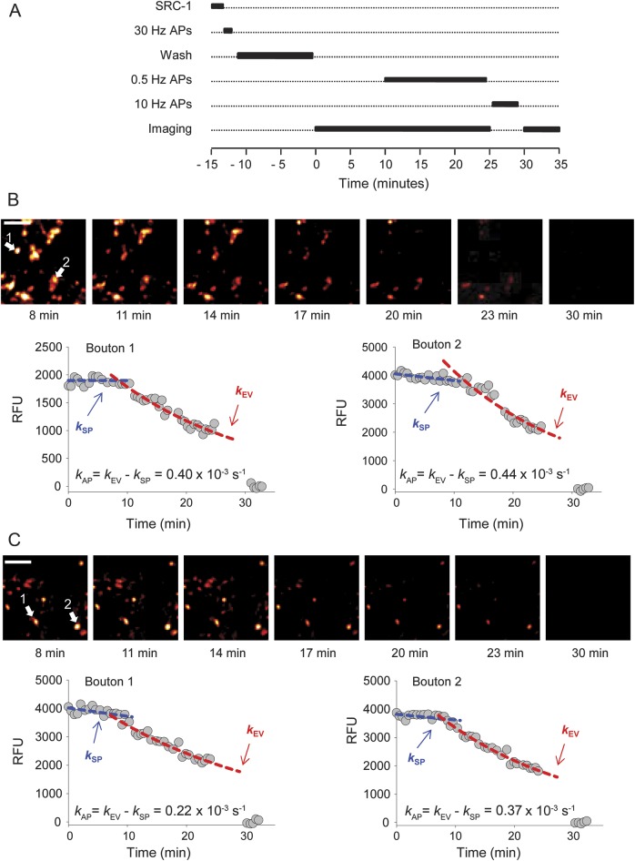
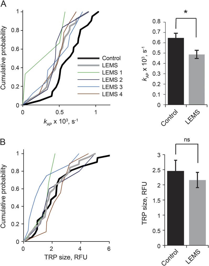
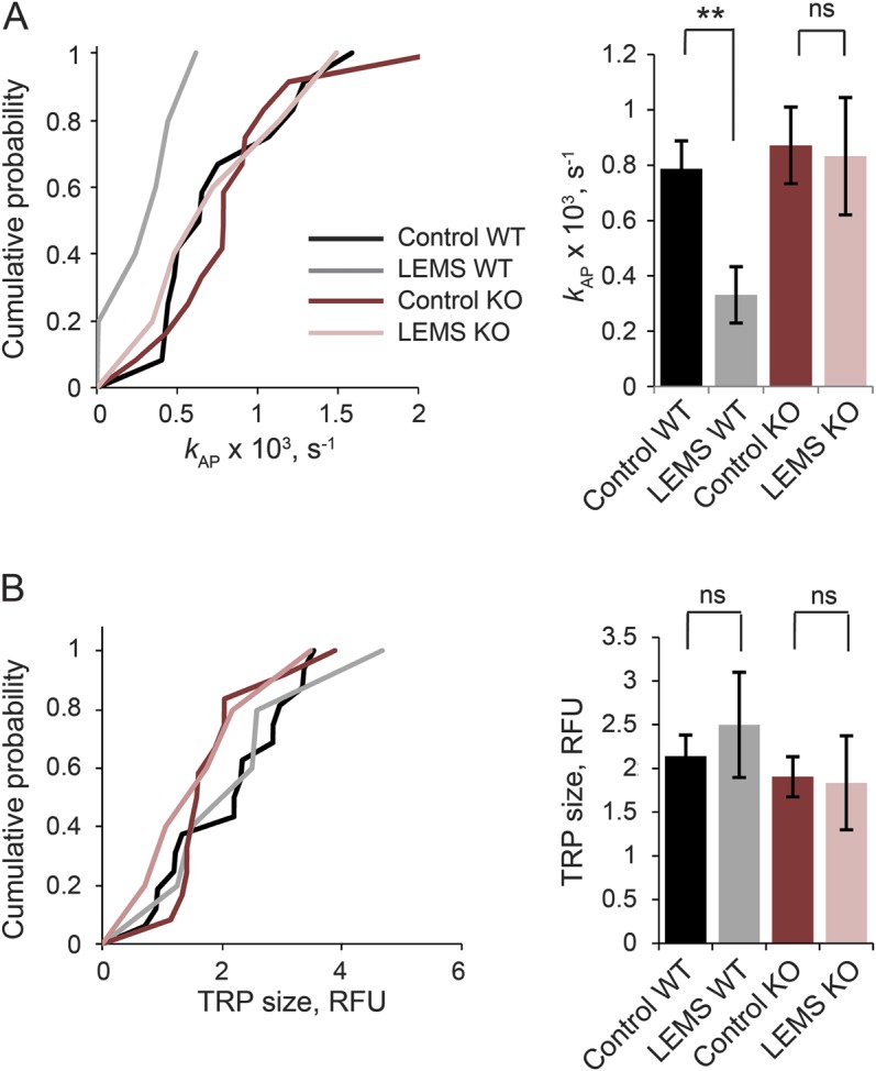
Methods: IgG was obtained from 4 patients with LEMS (3 males, 1 female), including 2 patients with lung malignancy. Antibodies against P/Q-type VGCCs were detected in all 4 patients, and against N-type VGCCs in 2. We incubated neuronal cultures with LEMS IgG and determined the size of the total recycling pool of synaptic vesicles and the rate of action potential–evoked exocytosis using fluorescence imaging of the amphiphilic dye SynaptoRed C1. Pooled IgG from healthy volunteers was used as a control. We repeated the experiments on synapses lacking P/Q-type calcium channels from a Cacna1a knockout mouse to determine whether these channels account for the pathogenic effect of LEMS IgG.
Results: LEMS IgG had no effect on the total recycling pool size but significantly reduced the rate of action potential–evoked synaptic exocytosis in wild-type neurons when compared with neurons treated with control IgG. In contrast, LEMS IgG had no effect on the rate of synaptic vesicle exocytosis in neurons lacking P/Q-type channels.
Conclusions: These data provide direct evidence that LEMS IgG inhibits neurotransmitter release by acting on P/Q-type VGCCs.
Figures
References
-
- Lennon VA, Kryzer TJ, Griesmann GE, et al. Calcium-channel antibodies in the Lambert-Eaton syndrome and other paraneoplastic syndromes. N Engl J Med 1995;332:1467–1474. 10.1056/NEJM199506013322203. - DOI - PubMed
-
- Motomura M, Lang B, Johnston I, Palace J, Vincent A, Newsom-Davis J. Incidence of serum anti-P/O-type and anti-N-type calcium channel autoantibodies in the Lambert-Eaton myasthenic syndrome. J Neurol Sci 1997;147:35–42. - PubMed
-
- Protti DA, Uchitel OD. Transmitter release and presynaptic Ca2+ currents blocked by the spider toxin omega-Aga-IVA. Neuroreport 1993;5:333–336. - PubMed
-
- Titulaer MJ, Lang B, Verschuuren JJ. Lambert-Eaton myasthenic syndrome: from clinical characteristics to therapeutic strategies. Lancet Neurol 2011;10:1098–1107. 10.1016/S1474-4422(11)70245-9. - DOI - PubMed
-
- Lang B, Newsom-Davis J, Wray D, Vincent A, Murray N. Autoimmune aetiology for myasthenic (Eaton-Lambert) syndrome. Lancet 1981;2:224–226. - PubMed
Publication types
MeSH terms
Substances
Grants and funding
LinkOut - more resources
Full Text Sources
Miscellaneous