. 2015 Jan 14;35(2):583-98.
doi: 10.1523/JNEUROSCI.2439-14.2015.

DAMP signaling is a key pathway inducing immune modulation after brain injury

Affiliations

DAMP signaling is a key pathway inducing immune modulation after brain injury

Arthur Liesz et al. J Neurosci. .

Erratum in

Abstract

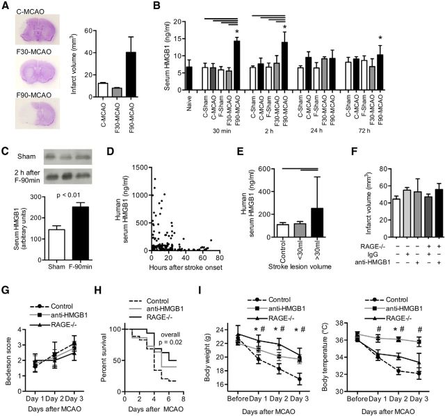
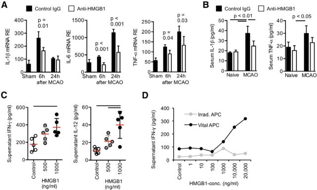
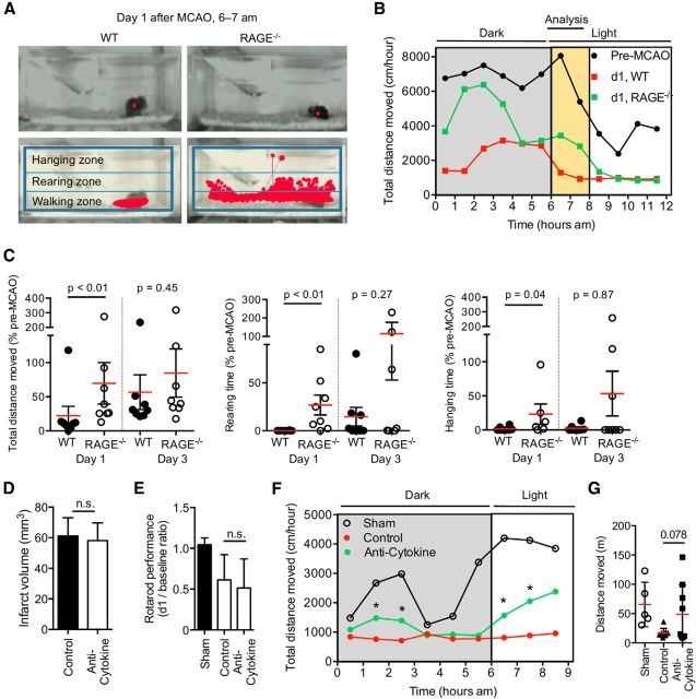
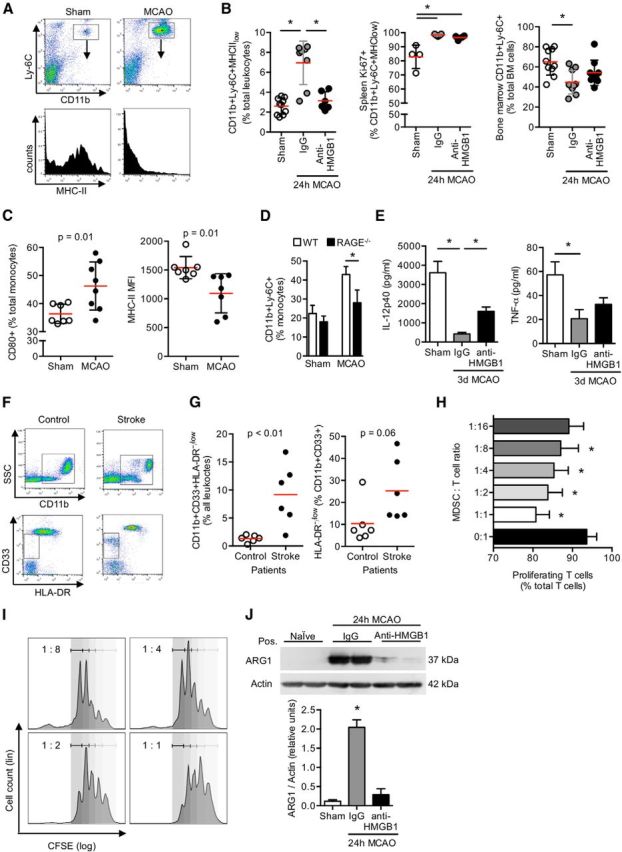
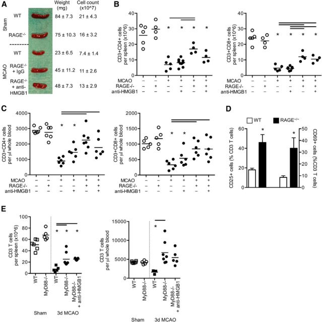
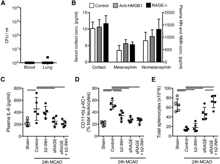
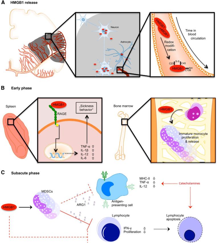
Acute brain lesions induce profound alterations of the peripheral immune response comprising the opposing phenomena of early immune activation and subsequent immunosuppression. The mechanisms underlying this brain-immune signaling are largely unknown. We used animal models for experimental brain ischemia as a paradigm of acute brain lesions and additionally investigated a large cohort of stroke patients. We analyzed release of HMGB1 isoforms by mass spectrometry and investigated its inflammatory potency and signaling pathways by immunological in vivo and in vitro techniques. Features of the complex behavioral sickness behavior syndrome were characterized by homecage behavior analysis. HMGB1 downstream signaling, particularly with RAGE, was studied in various transgenic animal models and by pharmacological blockade. Our results indicate that the cytokine-inducing, fully reduced isoform of HMGB1 was released from the ischemic brain in the hyperacute phase of stroke in mice and patients. Cytokines secreted in the periphery in response to brain injury induced sickness behavior, which could be abrogated by inhibition of the HMGB1-RAGE pathway or direct cytokine neutralization. Subsequently, HMGB1-release induced bone marrow egress and splenic proliferation of bone marrow-derived suppressor cells, inhibiting the adaptive immune responses in vivo and vitro. Furthermore, HMGB1-RAGE signaling resulted in functional exhaustion of mature monocytes and lymphopenia, the hallmarks of immune suppression after extensive ischemia. This study introduces the HMGB1-RAGE-mediated pathway as a key mechanism explaining the complex postischemic brain-immune interactions.

Keywords: HMGB1; RAGE; alarmins; immunomodulation; myeloid-derived suppressor cell; stroke.


Figures

Figure 1.
Figure 1.
Inhibition of the HMGB1 pathway after stroke improves mortality. A, Representative cresyl-violet-stained brain sections (left) after permanent distal MCA occlusion (C-MCAO), 30 min or 90 min transient proximal filament-MCAO (F30-MCAO and F90-MCAO), and analysis of mean infarct volumes in the respective MCAO model (3 d after MCAO, n = 12–15 per model, C57BL/6J WT mice). B, Serum HMGB1 concentration was measured by ELISA in unoperated naive animals, in the three stroke models, and in respective sham-operated animals (C-sham and F-sham, respectively) at the indicated time points after MCAO (n = 6 per group). *p < 0.05, between naive and the indicated group. Bars represent p < 0.05 between marked groups within one time point. Data are representative for 3 individual experiments per time point. C, Increase of serum HMGB1 in the 90 min filament-occlusion model was verified by Western blot analysis at 2 h after MCAO compared with the sham-operated control mice (n = 4 per group). D, Dot plot analysis of HMGB1 serum concentrations of ischemic stroke patients up to 72 h after stroke onset (n = 153). E, Serum HMGB1 was measured in matched control patients without stroke (n = 20) and in stroke patients. For analysis of stroke patients (n = 104), only blood samples of individual patients (no repetitive measurements) up to 24 h after symptom onset were included and patient groups dichotomized into small/moderate (<30 ml) and severe (>30 ml) lesion volumes. F, Stroke lesion volume was analyzed 3 d after 90 min filament-occlusion MCAO (F-90 min) in control animals (C57BL/6J) and RAGE−/− animals receiving anti-HMGB1 or control (isotype IgG) treatment. Infarct volumes did not significantly differ among groups (n = 18–34 per group, 4 individual experiments, p = 0.21). G, The Bederson score reflecting poststroke motor deficits was not affected by anti-HMGB1 treatment or RAGE deficiency. H, Kaplan–Meier curves of poststroke survival within the first week after F-90 min MCAO in control, anti-HMGB1 treated, and RAGE−/− mice. Overall p value = 0.02: Mantel-Cox test; n = 32(control), 17(RAGE−/−), 22(anti-HMGB1). p value (RAGE−/− vs control) = 0.008. I, Body weight (left) and rectal body temperature (right) were measured before surgery and on poststroke days 1–3 (n = 15–18 per group). *p < 0.05, control versus RAGE−/−. #p < 0.05, control versus anti-HMGB1. F–I, Data are representative of 4 individual experiments.
Figure 3.
Figure 3.
HMGB1 signaling induces early innate and adaptive immune activation. A, Cytokine mRNA expression was analyzed in spleens at the indicated time points after MCAO (n = 7 per group and time point). B, Expression levels of IL-1β and TNF-α were verified on protein level in the serum of control mice (isotype IgG) and anti-HMGB1-treated mice without brain lesion (naive) and 24 h after F-90 min MCAO by ELISA (n = 6 per group). A, B, Data are representative of 2 individual experiments per time point. C, The cytokine-inducing function of HMGB1 was tested in vitro. Interferon-γ (left) and IL-12 (right) concentration was analyzed in supernatants of mixed splenocyte cultures stimulated with disulphide-HMGB1 at the indicated concentration and compared with unstimulated samples (control). Bars represent p < 0.05 between indicated groups. Each data point represents one individual experiment. D, Naive CD3+ T cells were cocultured with untreated (vital) or irradiated APCs and stimulated with HMGB1 at the indicated concentration for 24 h. Interferon-γ concentrations were measured in the supernatant by ELISA (data representative of 5 individual experiments). Vital APCs are required for stimulation of IFN-γ production by T cells.
Figure 4.
Figure 4.
RAGE signaling mediates sickness-like behavior in the acute phase after stroke. A, Representative images of the unmodified tracking zones for continuous recording by the homecage behavior system (top). Analysis fields (hanging and rearing zones) and tracking (red lines) of mice in their home cages, indicated for 1 h recording (bottom). B, Brain ischemia by F-90 min MCAO induces a disturbed circadian rhythm with reduced activation response after a shift of night-day cycle at 6 A.M. Data are depicted for total distance moved per 1 h during the 12 h recording period (6 h dark and light cycle, each) and indicate a different degree of disturbed circadian rhythm between RAGE−/− and WT mice at day 1 (d1) after MCAO. C, Results of the homecage behavior analyses for overall mobility (moving distance: left) or motivation to challenging physical activity (rearing time: middle; hanging time: right) shown as cumulative data during the 2 h period from 6 A.M. to 8 A.M. at day 1 and day 3 after MCAO in WT control and RAGE−/− mice. Data were obtained in 3 individual experiments. D, Infarct volumetry 24 h after 90 min transient MCAO in mice receiving neutralizing antibodies against IL-1, TNF-α, and IL-6 (anti-cytokine) or isotype control IgG (control). E, Sensorimotor deficits were determined in the corresponding groups as in D and in a sham-operated group 24 h after F-90 min MCAO and normalized to baseline values. n.s., Not significant. F, The disturbed circadian rhythm after MCAO compared with sham treatment is improved in the anti-cytokine treated group as depicted by the approximation of the activity pattern to one of mice with sham surgery. G, Analysis of the total distance moved within the first 2 h of the light period (6 A.M. to 8 A.M.). D–G, Data were obtained from 3 individual experiments; n(sham) = 9, n(control) = 9, n(anti-cytokine) = 9.
Figure 5.
Figure 5.
Stroke induces immune exhaustion and expansion of immature monocytes via HMGB1-RAGE signaling in the subacute phase after stroke. A, Representative FACS plots for CD11b/Ly-6C expression of splenic mononuclear cells (CD45+ gated) 3 d after sham or F-90 min MCAO operation and the respective histogram for MHC-II expression of the double-positive cell population. B, Cell count of myeloid-derived progenitor cells (CD11+Ly-6C+MHC-IIlow) and their proliferation (CD11b+Ly-6C+Ki-67+) was analyzed in spleens at 24 h after sham operation or F90 min-MCAO with control (IgG) or anti-HMGB1 treatment (two left panels). Cell count of CD11b+Ly-6C+ cells in the bone marrow was determined in the same groups (right panel). Bars represent p < 0.05 between indicated groups. C, Splenic CD11c+ dendritic cells were analyzed for CD80 and MHC-II expression at 3 d after sham operation (control) or MCAO. D, Frequency of immature CD11+Ly-6C+ monocytes was compared between WT and RAGE−/− mice 3 d after F90 min-MCAO or sham surgery (n = 6 per group). E, Splenic CD11c+ dendritic cells (1 × 10⁵ cells) harvested from sham-operated animals and control antibody (IgG) or anti-HMGB1 treated animals 3 d after MCAO were stimulated with 1 μm CpG-ODN overnight, and IL12p40 and TNF-α cytokine concentrations were measured in the cell culture supernatant (n = 6 per group). B–E, Results are representative of at least two individual experiments per set. F, Representative FACS dot plots for CD11b (top) and CD33/HLA-DR expression (gated for CD11b+, bottom) of total blood leukocytes (CD45+ gated) from control or stroke patients. G, Blood cell counts of immature CD11b+CD33+monocytes (left) and their HLA-DR expression were analyzed in control patients with no neurological disease or in stroke patients at 24 h after extensive brain ischemia. H, Suppression assay analysis was performed to determine the suppressive function of splenic CD11b+Ly-6C+ monocytes harvested from donor animals 24 h after F90 min-MCAO (MDSC). T cells and MDSCs were cocultured at the indicated ratio and the proliferation of CFSE-labeled T cells analyzed by FACS (shown as percentage of total CD3+ T cells). CD11b+Ly-6C cells after MCAO are potent suppressors of T-cell proliferation. Data are representative of 5 individual experiments. I, Representative plots with fitted peaks for cell proliferation analysis by FlowJo software as used for MDSC suppression assay analysis shown before. Labeling in the plot indicates MDSC:T-cell ratio. J, Representative Western blot images (top) and analysis of ARG1 protein expression (bottom). ARG1 expression was measured in isolated splenic CD11b+ cells from sham-operated animals and control antibody (IgG) or anti-HMGB1 treated mice 24 h after F90 min-MCAO. Analysis is depicted as normalized ARG1 density values to actin expression. *p < 0.05 (n = 5 per group, 2 individual experiments).
Figure 6.
Figure 6.
HMGB1-RAGE signaling induces profound compromise of the peripheral adaptive immune system in the subacute phase after stroke. A, Representative images, mean weight, and cellularity (±SD) of spleens 3 d after sham operation or F90 min-MCAO in WT mice and RAGE−/− mice receiving control or anti-HMGB1 treatment (n = 5–7 per group). MCAO induces a profound reduction in spleen weight and cell count, which is significantly reversed in RAGE−/− mice. B, Flow cytometric analysis of absolute cell counts of Thelper (CD3+CD4+), Tcytotoxic (CD3+CD8+) cells per spleen and (C) per microliter of whole blood in sham-operated animals and 3 d after F90 min-MCAO in WT and RAGE−/− mice receiving additional control IgG antibodies or anti-HMGB1 treatment. *p < 0.05, between control animals of the same mouse strain and the indicated group. Bars represent p < 0.05 between marked groups within the MCAO+ groups. D, T-cell activation was analyzed (CD69+ right y-axis and CD25+ left y-axis, respectively) in spleens of WT and RAGE−/− mice 3 d after MCAO (n = 6 per group). E, The downstream signaling pathway of TLRs on CD3+ T cells was analyzed in MyD88−/− mice. T-cell counts were measured per total spleen (left) and microliters of whole blood (right) in sham-operated animals and 3 d after F90 min-MCAO in WT and MyD88−/− mice treated with control or anti-HMGB1 antibodies. *p < 0.05, between sham-operated animals of the same mouse strain and the indicated MCAO group. Bars represent p < 0.05 between marked groups within the MCAO groups.
Figure 7.
Figure 7.
The HMGB1-RAGE pathway is distinct from infectious or catecholamine-mediated effects. A, Microbiological analysis of blood samples and lung homogenates 3 d after 90 min Filament-MCAO for presence of bacteria. No significant growth of colony forming units (CFU) was detected after 24 h of incubation time on blood agar plates in any sample (n = 21, 2 individual experiments). B, Serum concentrations of cortisol and the catecholamines metanephrine and normetanephrine were measured 24 h after stroke induction (n = 7 per group, 2 individual experiments). Mice were treated with the β2-adrenoreceptor inhibitor ICI 118,551 (b2-INH) and/or sRAGE as indicated in Materials and Methods. C, sRAGE treatment, but not b2-INH, significantly reduced plasma IL-6 concentrations 24 h after F90 min-MCAO. D, The expansion of CD11b+Ly-6C MDSCs at 24 h after F90 min-MCAO was significantly reduced by sRAGE and, to a lesser extent, by b2-INH and without a measurable additive effect. E, The reduction in spleen cellularity induced by MCAO was abrogated by b2-INH as well sRAGE treatment with an additive effect. C–E, Data were obtained in three individual experiments; n = 5 per group.
Figure 8.
Figure 8.
Schematic overview of the proposed mechanisms of HMGB1 release and its biological activity after acute stroke. A, After acute vessel occlusion, cells of the hypoperfused territory become necrotic and release HMGB1 from its nuclear localization. Between 2 and 24 h after ischemia onset, the (C23-C45) disulphide “cytokine HMGB1” becomes the predominant HMGB1 isoform. B, Differential effects of HMGB1 in the spleen (left) and bone marrow (right) in the acute phase (<24 h) after stroke. Splenic monocytes are activated via a RAGE-dependent pathway, producing increased amounts of proinflammatory cytokines. In the bone marrow, HMGB1 induces proliferation of immature monocytes and their release to the circulation. C, The expanded population of (bone marrow-derived) immature monocytes has features of myeloid-derived suppressor cells, which inhibit lymphocyte activation via arginase (ARG1) secretion and might induce lymphocyte apoptosis. Splenic and circulating APCs have an “exhausted” phenotype characterized by reduced MHC-II expression and decreased cytokine production upon stimulation. Inadequate costimualtory signaling to lymphocyte induces T-cell dysfunction and might lead to apoptosis. Additionally, increased catecholamine levels in the subacute phase after stroke result in impaired monocyte function and lymphocyte reduction probably independent of HMGB1-RAGE signaling.

References

    1. Adachi O, Kawai T, Takeda K, Matsumoto M, Tsutsui H, Sakagami M, Nakanishi K, Akira S. Targeted disruption of the MyD88 gene results in loss of IL-1- and IL-18-mediated function. Immunity. 1998;9:143–150. doi: 10.1016/S1074-7613(00)80596-8. - DOI - PubMed
    1. An C, Shi Y, Li P, Hu X, Gan Y, Stetler RA, Leak RK, Gao Y, Sun BL, Zheng P, Chen J. Molecular dialogs between the ischemic brain and the peripheral immune system: dualistic roles in injury and repair. Prog Neurobiol. 2014;115:6–24. doi: 10.1016/j.pneurobio.2013.12.002. - DOI - PMC - PubMed
    1. Andrassy M, Volz HC, Igwe JC, Funke B, Eichberger SN, Kaya Z, Buss S, Autschbach F, Pleger ST, Lukic IK, Bea F, Hardt SE, Humpert PM, Bianchi ME, Mairbäurl H, Nawroth PP, Remppis A, Katus HA, Bierhaus A. High-mobility group box-1 in ischemia-reperfusion injury of the heart. Circulation. 2008;117:3216–3226. doi: 10.1161/CIRCULATIONAHA.108.769331. - DOI - PubMed
    1. Bederson JB, Pitts LH, Tsuji M, Nishimura MC, Davis RL, Bartkowski H. Rat middle cerebral artery occlusion: evaluation of the model and development of a neurologic examination. Stroke. 1986;17:472–476. doi: 10.1161/01.STR.17.3.472. - DOI - PubMed
    1. Bianchi ME. HMGB1 loves company. J Leukoc Biol. 2009;86:573–576. doi: 10.1189/jlb.1008585. - DOI - PubMed

Publication types

MeSH terms