. 2015 Jan-Mar;30(1):31-8.
doi: 10.4103/0972-3919.147532.

Potential role of (18)F-2-fluoro-2-deoxy-glucose positron emission tomography/computed tomography imaging in patients presenting with generalized lymphadenopathy

Affiliations

Potential role of (18)F-2-fluoro-2-deoxy-glucose positron emission tomography/computed tomography imaging in patients presenting with generalized lymphadenopathy

Sellam Karunanithi et al. Indian J Nucl Med. 2015 Jan-Mar.

Abstract

Generalized lymphadenopathy is a common and often vexing clinical problem caused by various inflammatory, infective and malignant diseases. We aimed to review briefly and highlight the potential role of (18)F-2-fluoro-2-deoxy-glucose ((18)F-FDG) positron emission tomography/computed tomography (PET/CT) in such patients. (18)F-FDG PET/CT can play an important role in the management of generalized lymphadenopathy. It can help in making an etiological diagnosis; can detect extranodal sites of involvement and employed for monitoring response to therapy.

Keywords: 18F-2-fluoro-2-deoxy-glucose positron emission tomography/computed tomography; generalized lymphadenopathy; lymphoma; sarcoidosis; tuberculosis.


Conflict of interest statement

Conflict of Interest: None declared.

Figures

Figure 1
Figure 1
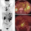
(a-c) A 58-year-old female with histologically confirmed Rosai Dorfman disease. Maximum intensity projection positron emission tomography (PET) image (a) and trans-axial PET/computed tomography images of neck (b), and abdomen (c), revealed multiple enlarged 18F-2-fluoro-2-deoxy-glucose avid bilateral cervical, axillary, retroperitoneal and mesenteric lymph nodes (arrows) along with splenic lesion (broken arrow)
Figure 2
Figure 2
(a-c) An 18-year-old male with Castleman's disease. Maximum intensity projection positron emission tomography (PET) image (a) revealed multiple enlarged 18F-2-fluoro-2-deoxy-glucose avid cervical, axillary, mediastinal, retroperitoneal and inguinal lymph nodes (arrows). Transaxial PET/computed tomography images demonstrating splenic (b, arrowhead) and bone marrow (c, broken arrow) involvement
Figure 3
Figure 3
(a-c) A 31-year-old female with sarcoidosis, for baseline evaluation. Maximum intensity projection positron emission tomography (PET) image (a) revealed multiple enlarged 18F-2-fluoro-2-deoxy-glucose (18F-FDG) avid cervical, axillary, mediastinal, portal, retroperitoneal and inguinal lymph nodes (broken arrows). Note is made of the “lambda sign” due to typical symmetrical 18F-FDG avid mediastinal lymph nodes involvement (a, bold arrows) transaxial PET/computed tomography images demonstrating lung (b, arrows) and splenic (c, arrows) involvement
Figure 4
Figure 4
(a-c) A 21-year-old female, known case of tuberculosis. Maximum intensity projection positron emission tomography (PET) image (a) revealed multiple enlarged 18F-2-fluoro-2-deoxy-glucose (18F-FDG) avid cervical, axillary, mediastinal, mesenteric, retroperitoneal and inguinal lymph nodes (arrows) transaxial PET/computed tomography (CT) images of thorax (b) 18F-FDG avid nodal (arrow) involvement. Note the calcification in mediastinal nodes (broken arrow). Transaxial PET/CT image of the lungs (c) 18F-FDG avid pulmonary involvement
Figure 5
Figure 5
(a-d) A 27-year-old male who presented with generalized lymphadenopathy. Transaxial positron emission tomography/computed tomography images (a-d) revealed mildly 18F-2-fluoro-2-deoxy-glucose avid bilateral cervical, axillary, mediastinal and abdominal lymph nodes (arrows). Biopsy from the lymph node was positive for actinomycosis
Figure 6
Figure 6
(a-f) A 38-year-old male with Hodgkin lymphoma (a and b), revealing 18F-2-fluoro-2-deoxy-glucose (18F-FDG) avid cervical, mediastinal, abdominal and inguinal lymph nodes, (a) apart from splenic (b, arrow) and liver (b, broken arrow) involvement. 34-year-old female with DLBCL (c and d), revealing 18F-FDG avid enlarged cervical, axillary, mediastinal, abdominal and inguinal lymph nodes, apart from splenic (c, broken arrow) and bone marrow (d, arrow) involvement. 73-year-old female with B-cell SLL (e and f), revealing mildly 18F-FDG-avid generalized lymph node involvement (arrows) with loss of normal architecture (f, arrows)
Figure 7
Figure 7
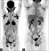
(a and b) A 57-year-old male with CD3+ peripheral T-cell lymphoma. Maximum intensity projection (MIP) positron emission tomography (PET) image (a) revealing 18F-2-fluoro-2-deoxy-glucose (18F-FDG) avid bilateral multiple cervical, axillary, mediastinal, abdomino-pelvic and inguinal lymph nodes (broken arrows), 27-year-old male with angiolymphoblastic T-cell lymphoma. MIP PET image (b) revealing mild to moderately 18F-FDG avid bilateral multiple cervical, axillary, mediastinal, abdomino-pelvic and inguinal lymph nodes (arrows)
Figure 8
Figure 8
(a-c) A 67-year-old female with biopsy proven ovarian carcinoma. Maximum intensity projection positron emission tomography (PET) image (a) and transaxial PET/computed tomography images (b and c) revealed multiple enlarged 18F-2-fluoro-2-deoxy-glucose avid cervical, axillary, mediastinal, retroperitoneal, mesenteric and pelvic metastatic lymph nodes (arrows)

References

    1. Foucar E, Rosai J, Dorfman R. Sinus histiocytosis with massive lymphadenopathy (Rosai-Dorfman disease): Review of the entity. Semin Diagn Pathol. 1990;7:19–73. - PubMed
    1. Karunanithi S, Singh H, Sharma P, Naswa N, Kumar R. 18F-FDG PET/CT imaging features of Rosai Dorfman disease: A rare cause of massive generalized lymphadenopathy. Clin Nucl Med. 2014;39:268–9. - PubMed
    1. Raveenthiran V, Dhanalakshmi M, Hayavadana Rao PV, Viswanathan P. Rosai-Dorfman disease: Report of a 3-year-old girl with critical review of treatment options. Eur J Pediatr Surg. 2003;13:350–4. - PubMed
    1. Deshayes E, Le Berre JP, Jouanneau E, Vasiljevic A, Raverot G, Seve P. 18F-FDG PET/CT findings in a patient with isolated intracranial Rosai-Dorfman disease. Clin Nucl Med. 2013;38:50–2. - PubMed
    1. Huang JY, Lu CC, Hsiao CH, Tzen KY. FDG PET/CT findings in purely cutaneous Rosai-Dorfman disease. Clin Nucl Med. 2011;36:13–5. - PubMed