. 2015;14(5):721-31.
doi: 10.1080/15384101.2014.995496.

New strategy for in vitro activation of primordial follicles with mTOR and PI3K stimulators

Affiliations

New strategy for in vitro activation of primordial follicles with mTOR and PI3K stimulators

Xinhui Sun et al. Cell Cycle. 2015.

Abstract

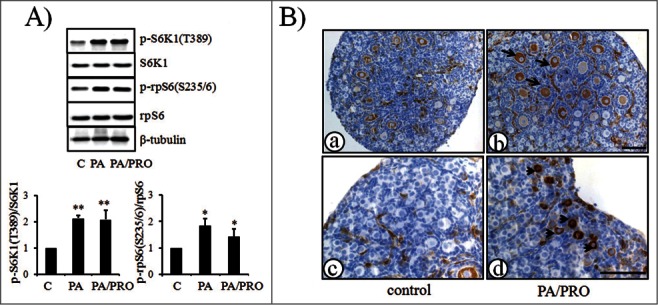
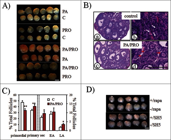
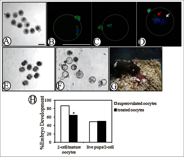
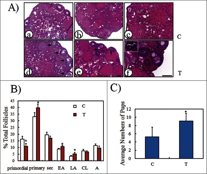
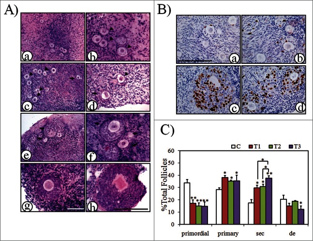
It had been known for decades that primordial follicles in mammalian ovaries are assembled with definite numbers and represent the ovarian reserve throughout the reproductive life. Intra-oocyte PI3K/mTOR pathways have been indicated to play a central role on the activation of primordial follicles. Genetic modified mouse models with chronic activation of PI3K/mTOR signals in primordial oocytes showed premature activation of all primordial follicles and eventually their exhaustion. On the other hand, this may suggest that, unlike chronic activation of PI3K/mTOR, its acute activation in infertility would activate primordial follicles, permitting fertility during the treatment. Previously, PI3K stimulators were reported as a temporary measure to accelerate primordial follicle activation and follicular development in both mouse and human, and were applied in the treatment of infertility in premature ovarian failure (POF) patients. To address whether mTOR stimulators could play similar role in the process, we transiently treated neonatal and aged mouse ovaries with mTOR stimulators-phosphatidic acid (PA) and propranolol. Our results demonstrated the stimulators increased activation of primordial follicles and the production of progeny. Human ovarian cortex cubes were also treated with mTOR or/and PI3K stimulators in vitro. When they were used separately, both of them showed similar promotive effects on primordial follicles. Surprisingly, after joint-treatment with the 2 kinds of stimulators together, synergistic effects on follicular development were observed. Based on increased efficiency of follicular activation in humans, here we propose in vitro transient treatment with mTOR and PI3K stimulators as an optimized protocol for the application in different clinical conditions with limited follicle reserve.

Keywords: PA, phosphatidic acid; POF, premature ovarian failure; PRO, propranolol.; PTEN, Phosphatase and Tensin Homolog deleted on chromosome 10; TSC1, tuberous sclerosis complex 1 or hamartin; TSC2, tuberous sclerosis complex 2 or tuberin; fertility preservation; follicular activation; mTOR, mammalian target of rapamycin; ovary; premature ovarian failure; primordial follicle.


Figures

Figure 1.
Figure 1.
Activation of mTOR-S6K1-rpS6 signaling pathway in neonatal ovaries after treatment with mTOR stimulators, PA and PRO. Ovaries were collected after 24 h of treatment. (A) Western blot of ovarian proteins with specific antibody for p-S6K1(T389), S6K1, p-rpS6(S235/6), rpS6 and β-tubulin. S6K1, rpS6, and β-tubulin were used as internal controls. (A,lower), Densitometry of western blot was quantified and shown by p-S6K1(T389)to S6K1 ratios and p-rpS6(S235/6)to rpS6 ratios. The ratios of p-S6K1(T389)to S6K1 and p-rpS6(S235/6)to rpS6 in control were designated as 1. The data in the graphs were presented as mean±SD of three independent experiments. *, P < 0.05; **, P < 0.01, compared with controls. (B) Immunostaining of p-rpS6 in ovaries treated w/o PA/PRO. (a) and (c), control non-treated ovaries. (b) and (d), PA/PRO treated ovaries. (c), (d) is the higher magnification of (a) and (b), which showed primordial follicle pools in the ovary. Black arrow, representative p-rpS6 signals in oocytes of growing secondary follicles; black arrowhead, representative p-rpS6 signals in oocytes of primordial follicles. Bar = 100 μm.
Figure 2.
Figure 2.
Increased ovarian development after PA and PRO stimulation. Paired neonatal ovaries were treated with or without mTOR stimulators, PA or PRO for 24h, followed by transplantation into kidney capsule of ovariectomized recipient mice. Transplanted ovarian grafts were collected 14 days later. (A) Isolated paired ovaries for PA (200 μM) vs. control; PRO (50 μM) vs. control; PA/PRO (200 μM/50 μM) vs. PA (200 μM); and PA/PRO (200 μM/50 μM) vs. PRO (50 μM). Bar = 1 mm. (B) ovarian histology by hematoxylin and eosin staining. (a) and (b), control ovaries; (c) and (d), PA/PRO treated ovaries. (b) and (d) are higher magnifications of (a) and (c), which showed clusters of primordial follicles (b, white arrows) in control ovaries and clusters of primary follicles (d, red arrows) in PA/PRO treated ovaries. Bar = 100 μm. (C) Distribution of follicles in ovaries treated with or without PA/PRO (n = 5). Data were shown with mean±SD. Sec, secondary follicle; EA, early antral follicle; LA, large antral follicle.*, P < 0.05; **, P < 0.01 vs. control. (D) Effects of PA plus PRO on ovarian development were blocked by mTOR inhibitor rapamycin (rapa) but not Akt inhibitor SH5. The symbol -/ indicated groups without the inhibitors. Bar = 1 mm.
Figure 3.
Figure 3.
Evaluation of oocyte quality by immunofluorescence, in vitro fertilization and early embryonic development. Day 3 ovaries were treated with PA (200 μM) and PRO (50 μM) for 24 h and transplanted into the kidney capsules of recipient mice for 18 days. Mature MII oocytes were retrieved 12 h later, after a single injection of hCG into recipient mice. (A) MII oocytes punctured from the transplanted ovaries. (B) Immunofluorescence of β-tubulin on spindle and first polar body of MII oocytes. Green, β-tubulin; blue, DNA. (C) and (D) Immunofluorescence of 5mC on (C) chromosomes of mature MII oocyte and (D) pronuclear of zygote. Zygotes with 2 pronucli were obtained 10h after in vitro fertilization. Red arrow, maternal pronuclear; white arrow, paternal pronuclear. Green, 5mC; Blue, DNA. (E) and (F) Early embryonic development of retrieved oocytes after in vitro fertilization. Representative figures for embryos reaching (E) 2-cell (24 h) and (F) blastocyst (96 h) stages. (G) Healthy pups with a host mother following 2-cell embryonic transfer. Mature MII oocytes retrieved from activated follicles were in vitro fertilized with donor sperm of the same strain and obtained 2-cell embryos were transferred into pseudopregnant ICR mice to establish pregnancy. (H) Efficiency of embryonic development. IVF rate (2-cell/mature oocytes) and successful 2-cell embryo transfer rate (live pups/2-cell) of mature oocytes retrieved from transplanted grafts were compared with those of mature oocytes from super-ovulated mice. *, P < 0.05 vs. superovulated oocytes. All bars = 100 μm.
Figure 4.
Figure 4.
Stimulation of follicular development in old mice by PA and PRO. PA/PRO (200 μM/50 μM) mixture was freshly prepared and injected directly into the ovarian bursa. For some animals, one lateral ovary was given PA/PRO and the other one received saline as control. Ovaries were collected 14 days later to evaluate follicular development. (A) Ovarian histology by hematoxylin and eosin staining. (a), (d) and (b), (e) show 2 pairs of representative ovaries. (a) and (b) are control ovaries with intra-bursal injection of saline and (d) and (e) are ovaries with intra-bursal injection of PA/PRO. Black arrows indicate developing antral follicles; (c) and (f) are higher magnifications of (b) and (e), respectively. The inset in (f) shows growing primary follicles. (B) Distribution of follicles in control and PA/PRO-treated ovaries (n = 6). Sec, secondary follicles; EA, early antral follicles; LA, large antral follicles; CL, corpus luteal; A, atresia. *, P < 0.05, as compared with controls. (C) Quantification of average pup numbers derived from old mice after bi-lateral intra-bursal injection of PA/PRO (T). Some animals served as controls and had only saline injection into bi-lateral ovarian bursa (C). Monitoring of the menstrual cycle began 18 days later and only the mice in estrous were mated, always with male mice of proven fertility. *, P < 0.05 vs. controls.
Figure 5.
Figure 5.
Activation of human primordial follicles by mTOR and/or PI3K stimulators. Human ovarian cortex cubes were divided into 4 groups and treated as the following: C, control; T1, mTOR stimulators, PA/PRO (200 μM/50 μM) incubation for 24 h; T2, PI3K stimulators, bpV/740Y-P (100 mM/250 μg/ml) for 24 h, 740Y-P(250 μg/ml) only for another 24 h; T3, mTOR and PI3K simulators, PA/PRO (200 μM/50 μM) for 24 h; bpV/740Y-P (100 μM/250 μg/ml) for another 24 h. After treatment, the tissues were continuously cultured in normal control media for another 4-5 days until a total 6 days of culture time. (A) Representative histology of human ovarian cortex cubes after 6 days of in vitro culture. Black arrow head, primordial follicles; black arrow, secondary follicles. (a) and (b), Ovarian cortex with primordial follicles before culture: (b) higher magnification of (a). (c) and (d), Follicles in control group after 6 days of culture: (c) primordial follicle cluster; (d) early secondary follicle. (e) and (f), Two representative clusters of growing secondary follicles in the group treated with mTOR stimulators (T1 group): (e) lower magnification; (f) higher magnification. (g) Representative secondary follicle with more than 3 layers of granulosa cells in PI3K stimulators treated group (T2 group). (h) Large secondary follicle after combined application of mTOR and PI3K stimulators (T3 group). (A, left), the same bar; (A, right), the same bar. Bars = 100 μm. (B) BrdU incorporation into granulosa cells of growing follicles. BrdU was added to culture media during the last 24h before collecting samples for fixation. (a) and (b), control group; (c) and (d), mTOR and PI3K stimulators joint-treated group (T3 group). Bar = 100 μm. (C) Follicle dynamics after treatment with or without mTOR and/or PI3K stimulators. Sec, secondary follicles; de, degenerated follicles. *, P < 0.05; **, P < 0.01 vs. control or as indicated.

References

    1. McLaughlin EA, McIver SC. Awakening the oocyte: controlling primordial follicle development. Reproduction 2009; 137:1-11; PMID:18827065; http://dx.doi.org/10.1530/REP-08-0118 - DOI - PubMed
    1. McGee EA, Hsueh AJ. Initial and cyclic recruitment of ovarian follicles. Endocr Rev 2000; 21:200-14; PMID:10782364 - PubMed
    1. Macklon NS, Fauser BC. Aspects of ovarian follicle development throughout life. Horm Res 1999; 52:161-70; PMID:10725781; http://dx.doi.org/10.1159/000023456 - DOI - PubMed
    1. Okeke T, Anyaehie U, Ezenyeaku C. Premature menopause. Ann Med Health Sci Res 2013; 3:90-5; PMID:23634337; http://dx.doi.org/10.4103/2141-9248.109458 - DOI - PMC - PubMed
    1. Goswami D, Conway GS. Premature ovarian failure. Hum Reprod Update 2005; 11:391-410; PMID:15919682; http://dx.doi.org/10.1093/humupd/dmi012 - DOI - PubMed

Publication types

MeSH terms