Review
. 2015;14(3):305-14.
doi: 10.4161/15384101.2014.991185.

Meiotic crossover patterns: obligatory crossover, interference and homeostasis in a single process

Affiliations
Review

Meiotic crossover patterns: obligatory crossover, interference and homeostasis in a single process

Shunxin Wang et al. Cell Cycle. 2015.

Abstract

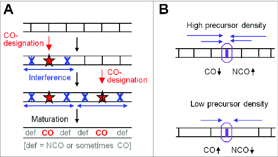
During meiosis, crossover recombination is tightly regulated. A spatial patterning phenomenon known as interference ensures that crossovers are well-spaced along the chromosomes. Additionally, every pair of homologs acquires at least one crossover. A third feature, crossover homeostasis, buffers the system such that the number of crossovers remains steady despite decreases or increases in the number of earlier recombinational interactions. Here we summarize recent work from our laboratory supporting the idea that all 3 of these aspects are intrinsic consequences of a single basic process and suggesting that the underlying logic of this process corresponds to that embodied in a particular (beam-film) model.

Keywords: BF, beam-film; CO, crossover; DDF, designation driving force; DSBs, double-strand breaks; NCO, noncrossover; SC, synaptonemal complex; STUbL, SUMO-targeted ubiquitin ligase; beam-film model; crossover; crossover homeostasis; crossover interference; meiosis; obligatory crossover; recombination.


Figures

Figure 1.
Figure 1.
Meiotic crossover (chiasma) patterns. (A) Meiosis comprises 2 successive rounds of chromosome segregation. At Meiosis I, homologous chromosomes (red and black) segregate to opposite poles. The segregating chromosomes are connected by the combined effects of a CO between one sister of each homolog and links between sister chromatids along their arms. Because of this connection, when homologous chromosomes are connected to opposite poles, tension arises on their corresponding centromeres (red and black circles). When all pairs are under such tension, cell cycle regulatory mechanisms license onset of Anaphase I. At Meiosis II, sister chromatids segregate to opposite poles, this time guided by connections between sister centromeres.(B) In many organisms, the CO-generated links between homologs at Meiosis I (A) are seen cytologically as "chiasmata." Note that in favorable cases, sister cohesion is disrupted around chiasma sites (arrows) in accord with the connection of non-sister chromatids by COs at those positions (from ref.4).
Figure 2.
Figure 2.
Even spacing of chiasmata and pachytene CO-correlated recombination complexes. (A) Pairs of homologous grasshopper chromosomes ("bivalents") linked by chiasmata (from ref.5). Note that each bivalent has at least one chiasma, sometimes only one (arrow) and that multiple chiasmata (arrow heads) on a single bivalent are spaced far away from one another. (B) A diplotene bivalent of Sordaria macrospora showing evenly-spaced chiasmata (D.Z.). (C) A pachytene bivalent of Sordaria macrospora. SC illuminated with Sme4-GFP (green) and decorated with evenly-spaced CO-correlated Hei10-T3-mCherry foci (red) (D.Z. unpublished) (D) A pachytene bivalent of budding yeast. SC (red) and CO-correlated Zip3 foci (green) illuminated with antibodies against Zip1 and Myc (for detection of Myc tagged Zip3) respectively (from ref.11). (E) Patterning along mammalian mitotic metaphase chromosomes as manifested in evenly-spaced alternating domains of TopoII and condensin I (from ref.76).
Figure 3.
Figure 3.
Crossover Interference in wild-type and mutant budding yeast. CoC relationships for positions of CO-correlated Zip3 foci along pachytene bivalents of budding yeast in wild type and mutant situations (from ref.11). (A) Experimental data for Chromosome XV (black) and BF best-fit simulation (green). Left: CoC relationships, plotted as function of inter-interval distance in μm axis length. Right: the distribution of Zip3 foci (COs) per bivalent. A convenient measure of the apparent strength of CO interference is provided by the inter-interval distance at which CoC = 0.5. In wild type, this "interference distance" is ∼300 nm. This value corresponds approximately to the average distance between adjacent COs. (B) Meiotic depletion of TopoII increases the CoC at smaller inter-interval distances and shifts the CoC curve to the left (pink) as compared to wild type [black; from (A)], with corresponding BF best fit simulation (right, green). Interference distance in the mutant is ∼200 nm. (C) A yeast condensin mutant has longer chromosome axes. CoC relationships in the mutant (red) are the same as in wild type (black) when the metric of inter-interval distance is physical chromosome length (μm) (top) and are different when the metric is genomic distance (Kb) (bottom). (D) Model for the role of TopoII in crossover interference. Chromatin expansion puts stress on the protein/protein/DNA meshwork of chromosome axes (top). Local CO-designation gives local relaxation, which then spreads along the axes; TopoII is required to adjust the relationships among DNA segments in response to this relaxation. All images are from ref.
Figure 4.
Figure 4.
The logic of beam-film model and CO homeostasis. (A) The general logic of the beam-film model (from ref.10). The "designation driving force" works on an array of precursors (vertical black lines) and promotes CO designation (red stars) with resulting spreading of the interference signal outward in both directions, dissipating with distance. Sequential CO designations with ensuing spreading interference signals lead to COs that tend to be evenly spaced (text). (B) Crossover homeostasis from the perspective of an individual DSB-mediated recombinational interaction according to the logic of the BF model. Crossover homeostasis results from interplay between precursor density and spreading interference. At high (low) precursor (vertical black lines) density, a precursor will be more (less) affected by the spreading interference signal (blue arrows) from nearby crossover-designations and thus will be less (more) likely to be a crossover.
Figure 5.
Figure 5.
Recombination occurs in the context of chromosome structural axes. (A) Meiotic prophase chromosomes comprise co-oriented sister linear arrays of chromatin loops (black and brown), the bases of which are decorated by structural components in a protein/DNA meshwork (blue and green) (ref.29) These proteins (e.g., Topoisomerase II, condensins, cohesins and meiotic specific Red1 and Hop1) bind to axis association sites, which are usually regularly distributed, locally AT-rich regions. (B) A model for recombinosome-mediated homolog juxtaposition (ref.1). (i) One dual loop (black and brown) from one homolog is tethered onto axis (green; axial meshwork not shown) by recombinosome complex (not shown) and a DSB forms (2 red arrow heads) within one chromatin loop (black). (ii) The "leading" end of the DSB is released and searches for a homologous sequence on its homolog partner while the "lagging" DSB end is retained on the axis. (iii) The homolog is caught and bought into closer proximity, to ∼400 nm (the equivalent stage in Panels C and D). (iv) Finally the 2 axes move closer, to a distance of ∼100 nm, and synaptonemal complex forms (not shown). (C, D) DSB-mediated inter-axis bridges along coaligned late leptotene axes in Sordaria macrospora. (C) In late leptotene nuclei, pairs of foci of meiotic helicase Mer3 occur along the axes of homologous chromosomes as illuminated with fluorescent axis component Spo76/Pds5 (from ref.35). Foci of each pair mark the 2 ends of a single DSB, implying that single DSB-mediated recombinational interaction is bridging the 2 axes. (D) Left: coaligned axes as in (C). Middle and Right: DAPI staining reveals inter-axis DNA bridges that presumptively correspond to the recombination-mediated bridges in (C) (D.Z.)

References

    1. Kleckner N, Zhang L, Weiner B, Zickler D. Meiotic chromosome dynamics. In: Rippe, ed. Genome Organization and Function in the Mammalian Cell Nucleus. New York: John Wiley and Sons, 2011:487-534.
    1. Zickler D, Kleckner N. Recombination, pairing and synapsis of homologs in meiosis. In: Kowalczykowski S, Hunter N, Heyer WD. DNA Replication. Cold Spring Harbor: Cold Spring Harbor Press, 2015; in press. - PMC - PubMed
    1. Berchowitz LE, Copenhaver GP. Genetic interference: don't stand so close to me. Curr Genomics 2010; 11(2):91-102; PMID:20885817; http://dx.doi.org/10.2174/138920210790886835 - DOI - PMC - PubMed
    1. Jones GH, Franklin FC. Meiotic crossing-over: obligation and interference. Cell 2006; 126(2):246-8; PMID:16873056; http://dx.doi.org/10.1016/j.cell.2006.07.010 - DOI - PubMed
    1. Jones GH. The control of chiasma distribution. Symp Soc Exp Biol 1984; 38:293-320; PMID:6545727 - PubMed

Publication types

LinkOut - more resources