. 2015 Jan 15;6(1):e1594.
doi: 10.1038/cddis.2014.552.

PKR downregulation prevents neurodegeneration and β-amyloid production in a thiamine-deficient model

Affiliations

PKR downregulation prevents neurodegeneration and β-amyloid production in a thiamine-deficient model

F Mouton-Liger et al. Cell Death Dis. .

Abstract

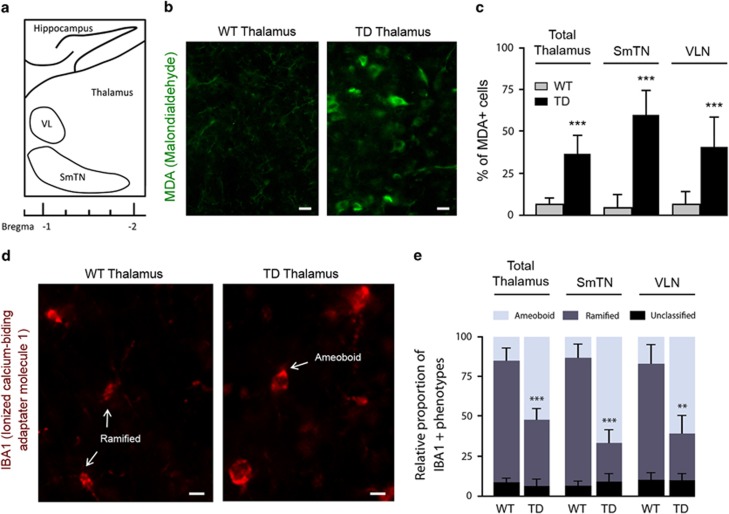
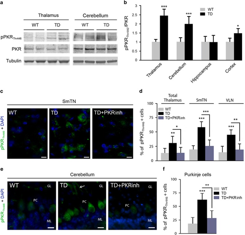
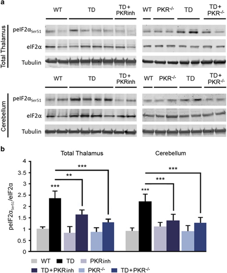
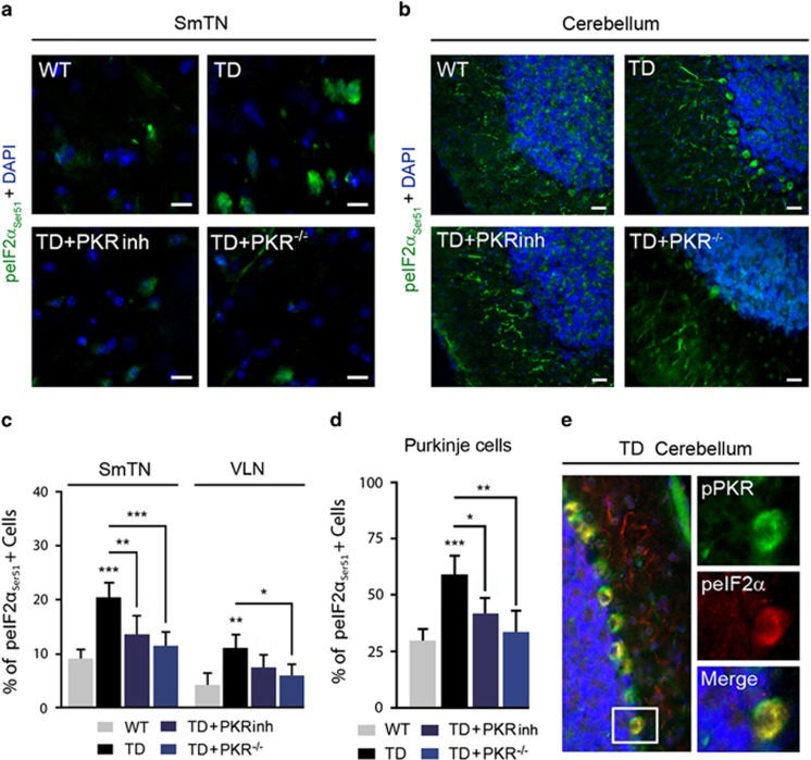
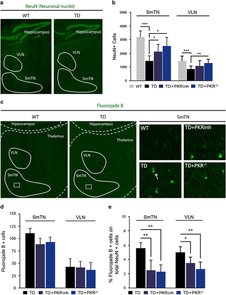
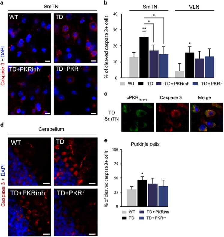
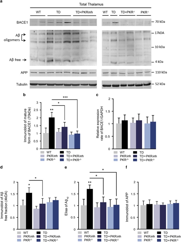
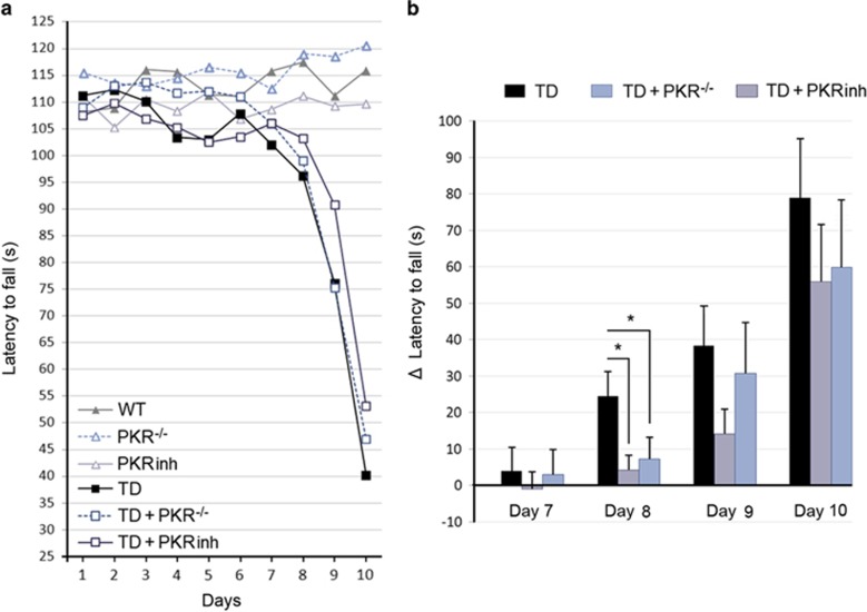
Brain thiamine homeostasis has an important role in energy metabolism and displays reduced activity in Alzheimer's disease (AD). Thiamine deficiency (TD) induces regionally specific neuronal death in the animal and human brains associated with a mild chronic impairment of oxidative metabolism. These features make the TD model amenable to investigate the cellular mechanisms of neurodegeneration. Once activated by various cellular stresses, including oxidative stress, PKR acts as a pro-apoptotic kinase and negatively controls the protein translation leading to an increase of BACE1 translation. In this study, we used a mouse TD model to assess the involvement of PKR in neuronal death and the molecular mechanisms of AD. Our results showed that the TD model activates the PKR-eIF2α pathway, increases the BACE1 expression levels of Aβ in specific thalamus nuclei and induces motor deficits and neurodegeneration. These effects are reversed by PKR downregulation (using a specific inhibitor or in PKR knockout mice).


Conflict of interest statement

Professor Jacques Hugon is a consultant for Roche, Sanofi, Novartis, Xigen, Eisai and Lundbeck. Dr. Claire Paquet is a consultant for Lilly and Novartis. Dr. Julien Dumurgier is a consultant for Novartis. Dr. Patrick Bernardelli, Dr. Véronique Taupin, Dr. Thomas Rooney and Dr. Laurent Pradier are fully employed by Sanofi. The remaining authors declare no conflict of interest.

Figures

Figure 1
Figure 1
Markers of oxidative stress (MDA) and microglial cell activation (IBA1) in the TD thalamus. (a) Schematic sagittal representation of thalamic nuclei distribution: SmTN; and VLN. (b) MDA immunohistochemistry (green) on the sagittal sections of WT and TD thalamus. (c) Percentage of MDA-positive cells in total thalamus, SmTN and VLN in WT (n=6) and TD mice (n=6). (d) IBA1 labeling (red) on the sagittal sections of WT and TD thalamus. (e) Quantification of relative proportion of IBA1-positive microglial cell phenotypes: ramified shape (inactivated state) and amoeboid shape (activated state) in WT (n=7) and TD mice (n=7). Unclassified shape corresponds to intermediate state or cells that were unable to be quantified. Scale bars: 10 μm. **P<0.01, ***P<0.001
Figure 2
Figure 2
Activation of PKR in TD brain studied by immunoblotting and immunohistochemistry in the thalamus and cerebellum. Immunoblot analyses (a) and corresponding quantification of pPKRThr446 and full PKR (b) in WT and TD mice protein samples, in the thalamus, cerebellum, hippocampus and cortex normalized on Tubulin levels. WT (n=14) and TD (n=14). *P<0.05, **P<0.01, ***P<0.001. (c) Immunohistochemistry (green) with a pPKRThr446 antibody on WT, TD and TD pretreated with PKRinh mice in sagittal sections of (c) SmTN and (e) cerebellum. (d) The positive cell counting confirmed that PKR is activated after TD treatment and its increase is attenuated by PKRinh administration in total thalamus, specifically in SmTN and VLN. (e) Cerebellar pPKRThr446 patterns in WT, TD and TD+PKRinh. pPKRThr446 staining is almost absent in granular layer (GL) and molecular layer (ML) but exhibits strong signals and increased after TD in Purkinje cells (PC), both in cell bodies and processes (arrow). (f) The percentages of PC with pPKRThr446 nuclear localization have been quantified, and as expected, pPKRThr446 staining is decreased in PC after PKRinh administration WT (n=8) and TD (n=8). Scale bars: 10 μm
Figure 3
Figure 3
PKR inhibition controls eIF2α activity in TD mouse thalamic and cerebellar neurons. (a and b) Immunoblot analyses (a) and corresponding quantification (b) after normalization on Tubulin of peIF2αSer51/eIF2α ratios in the three groups of mice non-exposed to TD (WT (n=14), PKRinh (n=10) and TD+PKR−/− (n=12)) and in the three groups of vitamin B1-deficient mice (TD (n=14), TD+PKRinh (n=10) and TD+PKR−/− (n=12)) in the total thalamus and cerebellum
Figure 4
Figure 4
eIF2αSer51 cellular localization in the thalamus and cerebellum in TD mice. (a and b) Double-labeling of DAPI (4,6-diamidino-2-phenylindole; blue) and peIF2αSer51 (green) in the (a) SmTN and (b) cerebellum of WT, TD, TD+PKRinh and TD+PKR−/− mice. peIF2αSer51 in cerebellum is present in Purkinje cells' cytoplasm and mainly increase after TD at the level of the cells bodies. Scale bars: 10 μm. (c and d) Quantification of positive neurons in SmTN and (c) VLN and (d) in Purkinje cells suggests that peIF2αSer51 phosphorylation is dependent of pPKRThr446 activation. *P<0.05, **P<0.01, ***P<0.001. (e) Confocal analysis showed a colocalization of pPKRThr446 (green) and peIF2α (red) in TD cerebellum of the cerebellum. Boxes represent higher magnification at the level of Purkinje cells, where colocalization (yellow) is mainly present in cell bodies
Figure 5
Figure 5
Regional TD-induced neuronal loss and neurodegeneration in the thalamus reduced by modulation of PKR activation. (a) Immunohistochemistry of neuronal nuclear marker NeuN (green) shows a decreased number of neuron in the TD thalamus compared with control mice, in particular in SmTN and VLN (surrounded areas). (b) The number of marked NeuN cells' evaluation indicates that TD-induced neuronal loss is reversed by PKR inhibition in the SmTN and in VLN. WT (n=8), TD (n=8) TD+PKRinh (n=8) and TD+PKR−/− (n=7). (c) F-J B histofluorescence in thalamic regions (surrounded nuclei: SmTN and VLN) reveals, as visible in higher magnification boxes, neurodegenerative neurons in TD mice. Note that WT thalamus did not present a positive signal. (d and e) Relative analysis as number of F-J B+ cells in SmTN and VLN (d) or the percent normalized on the mean number of NeuN+ (e). The percentage of TD-damaged neurons is decreased by PKR inhibition (TD+PKRinh and TD+PKR−/−). WT (n=8), TD (n=8) TD+PKRinh (n=8) and TD+PKR−/− (n=7); *P<0.05, **P<0.01, ***P<0.001
Figure 6
Figure 6
Immunolocalization of caspase 3 is enhanced in TD SmTN and Purkinje cells, and decreased by inhibition of PKR. (a) Double-labeling of nuclear marker DAPI (4,6-diamidino-2-phenylindole; blue) and caspase 3 (red) in SmTN of WT, TD, TD+PKRinh and TD+PKR−/− mice. (b) The percentage of cleaved-caspase 3-positive neurons increased by TD is significantly reduced after downregulating PKR in SmTN (left) but not in VLN (right). (TD+PKRinh and TD+PKR−/−). (c) Confocal microscopy of double staining of caspase 3 (red)/PKR (green) performed with the Zenon Labeling Kit on coronal sections of TD submedial nuclei of the thalamus (SmTN) showing cytoplasmic colocalizations in neurons (yellow). (d and e) Caspase 3 immunolocalization in Purkinje cell layers (cell bodies and arborization) is amplified in TD models but is not reversed by downregulation of PKR. WT (n=8), TD (n=8) TD+PKRinh (n=8) and TD+PKR−/− (n=7) *P<0.05, **P<0.01
Figure 7
Figure 7
Inhibition of PKR-eIF2α pathway by PKRinh or in PKR−/− mice decreases BACE1 levels and Aβ (Amyloid β) production. (a) Immunoblot on thalamus extracts and quantification of (b) BACE1, (d) Aβ oligomers and (f) APP. The blots show that TD induces BACE1 maturation and Aβ production without altering APP levels. PKR inhibition partially reduced the effects of TD. WT (n=8), PKRinh (n=8), PKR−/− (n=7) TD (n=8), TD+PKRinh (n=8) and TD+PKR−/− (n=7). (c) Transcriptional activity on BACE1 is not modified in the TD thalamus. Effect of TD on BACE1 mRNA levels is assessed with quantitative reverse transcriptase–PCR. BACE1 mRNA levels were normalized to GAPDH (glyceraldehyde 3-phosphate dehydrogenase) mRNA levels. WT (n=6), PKRinh (n=5), PKR−/− (n=5) TD (n=6), TD+PKRinh (n=5) and TD+PKR−/− (n=5). (e) The TD-dependent increase of Aβ1–42 peptide was confirmed by a sandwich enzyme-linked immunosorbent assay (ELISA) method and is expressed as means±S.E.M. Aβ1–42. The values from TD mice are expressed relative to the values from WT mice that were set to ‘1'. Immunoblots and ELISA were both performed on six different groups of mice: WT (n=8), PKRinh (n=8), PKR−/− (n=7) TD (n=8), TD+PKRinh (n=8), and TD+PKR−/− (n=7). *P<0.05, **P<0.01, ***P<0.001
Figure 8
Figure 8
The rotarod motor performance impaired in TD mice is rescued after PKRinh administration or in PKR−/− mice. (a) Diagram illustrating latency to fall measured on separate days reveals a strong decrease of motor performance in the three groups of TD mice. (b) Statistical analysis of measures for each animal of the delay between the rotarod performance between the first day of the experiment and on days 7, 8, 9 or 10. At the beginning of the decline (day 8), downregulation of PKR maintains motor performance of animals with TD. WT (n=14), PKRinh (n=14), PKR−/− (n=12) TD (n=14), TD+PKRinh (n=14) and TD+PKR−/− (n=12). *P<0.05

Similar articles

Cited by

References

    1. Baker KG, Harding AJ, Halliday GM, Kril JJ, Harper CG. Neuronal loss in functional zones of the cerebellum of chronic alcoholics with and without Wernicke's encephalopathy. Neuroscience 1999; 91: 429–438. - PubMed
    1. Chornyy S, Parkhomenko J, Chorna N. Thiamine deficiency caused by thiamine antagonists triggers upregulation of apoptosis inducing factor gene expression and leads to caspase 3-mediated apoptosis in neuronally differentiated rat PC-12 cells. Acta Biochim Pol 2007; 54: 315–322. - PMC - PubMed
    1. Calingasan NY, Huang PL, Chun HS, Fabian A, Gibson GE. Vascular factors are critical in selective neuronal loss in an animal model of impaired oxidative metabolism. J Neuropathol Exp Neurol 2000; 59: 207–217. - PubMed
    1. Karuppagounder SS, Shi Q, Xu H, Gibson GE. Changes in inflammatory processes associated with selective vulnerability following mild impairment of oxidative metabolism. Neurobiol Dis 2007; 26: 353–362. - PMC - PubMed
    1. Ke ZJ, Gibson GE. Selective response of various brain cell types during neurodegeneration induced by mild impairment of oxidative metabolism. Neurochem Int 2004; 45: 361–369. - PubMed

Publication types

MeSH terms

LinkOut - more resources