. 2015 Jan 15;6(1):e1596.
doi: 10.1038/cddis.2014.563.

Dose-dependent effects of selenite (Se(4+)) on arsenite (As(3+))-induced apoptosis and differentiation in acute promyelocytic leukemia cells

Affiliations

Dose-dependent effects of selenite (Se(4+)) on arsenite (As(3+))-induced apoptosis and differentiation in acute promyelocytic leukemia cells

S Wang et al. Cell Death Dis. .

Abstract

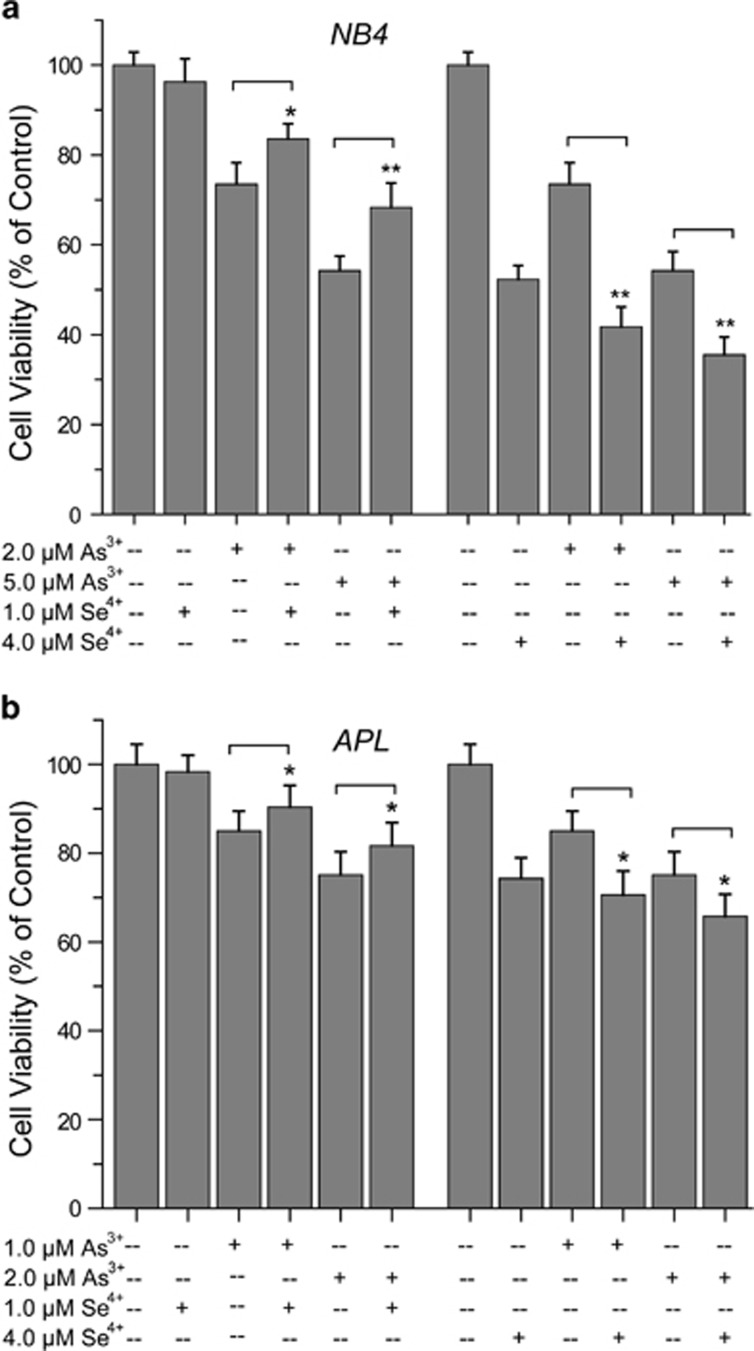
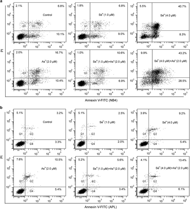
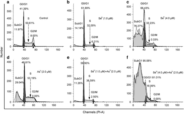
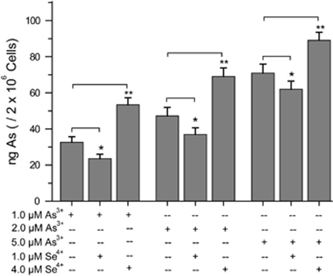
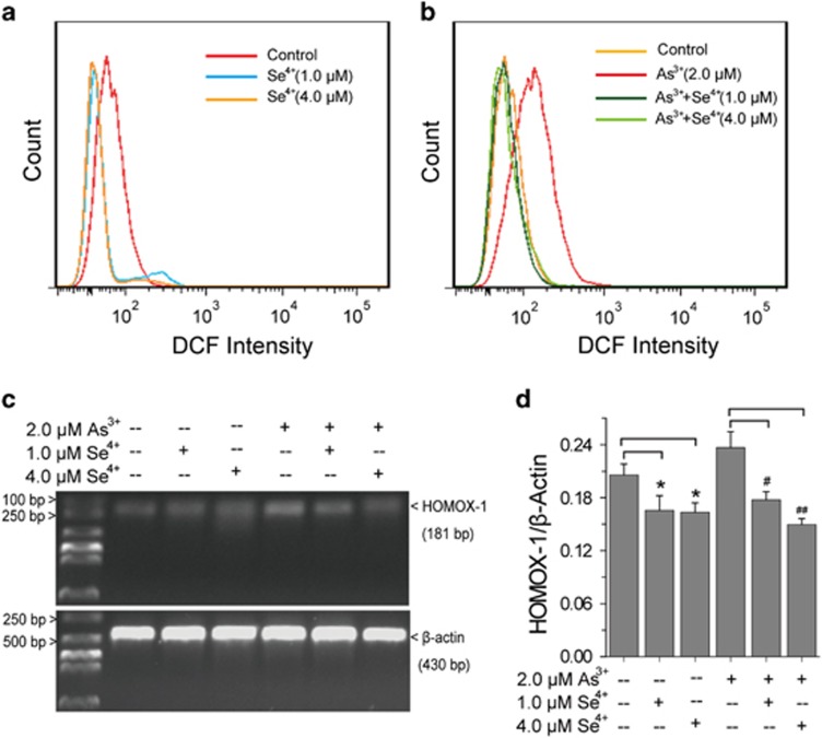
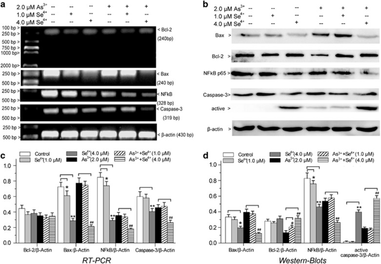
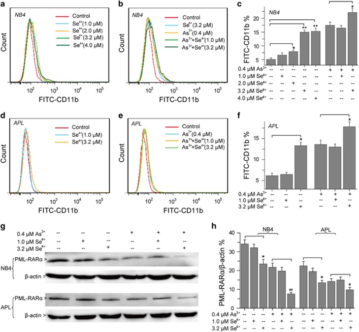
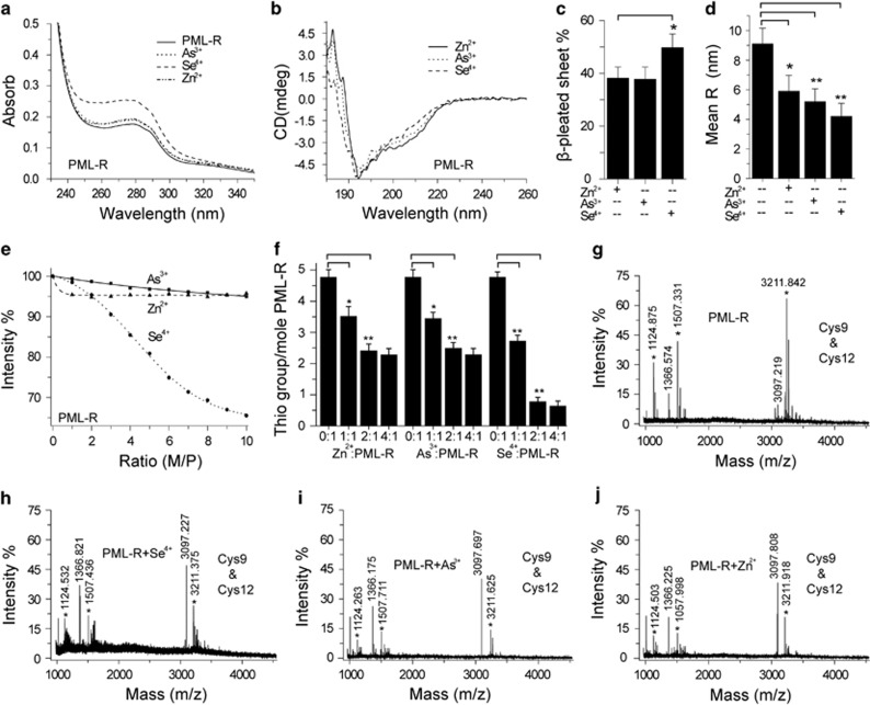
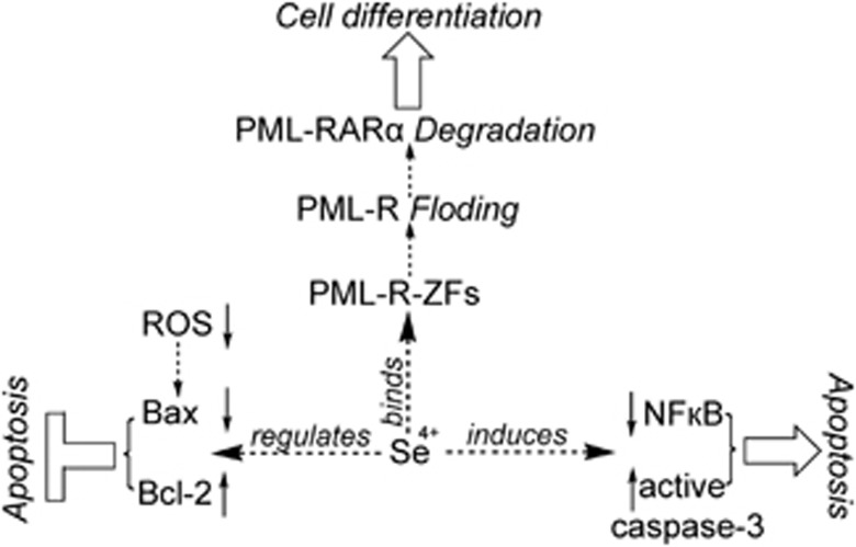
To enhance the therapeutic effects and decrease the adverse effects of arsenic on the treatment of acute promyelocytic leukemia, we investigated the co-effects of selenite (Se(4+)) and arsenite (As(3+)) on the apoptosis and differentiation of NB4 cells and primary APL cells. A 1.0-μM concentration of Se(4+) prevented the cells from undergoing As(3+)-induced apoptosis by inhibiting As(3+) uptake, eliminating As(3+)-generated reactive oxygen species, and repressing the mitochondria-mediated intrinsic apoptosis pathway. However, 4.0 μM Se(4+) exerted synergistic effects with As(3+) on cell apoptosis by promoting As(3+) uptake, downregulating nuclear factor-кB, and activating caspase-3. In addition to apoptosis, 1.0 and 3.2 μM Se(4+) showed contrasting effects on As(3+)-induced differentiation in NB4 cells and primary APL cells. The 3.2 μM Se(4+) enhanced As(3+)-induced differentiation by promoting the degradation of promyelocytic leukemia protein-retinoic acid receptor-α (PML-RARα) oncoprotein, but 1.0 μM Se(4+) did not have this effect. Based on mechanistic studies, Se(4+), which is similar to As(3+), might bind directly to Zn(2+)-binding sites of the PML RING domain, thus controlling the fate of PML-RARα oncoprotein.


Figures

Figure 1
Figure 1
Effects of Se4+ on As3+-induced cell death. (a) The viability of NB4 cells was determined with the WST-1 cell proliferation assay after 48 h of treatment. (b) The viability of primary APL cells. Error bars represent S.D. from the mean of three separate experiments. *P<0.05 and **P<0.01 compared with As3+-treated cells
Figure 2
Figure 2
Effects of As3+ and Se4+ on cell apoptosis. (a) Cell apoptosis in NB4 cells measured by Annexin V-FITC and PI double staining. (b) Cell apoptosis in primary APL cells. Q1 and Q3 respectively represent the proportions of dead cells and living cells, and Q2 and Q4 were used to calculate apoptotic cells. Figures show a representative experiment from three independent experiments
Figure 3
Figure 3
Cell cycle distribution profiles of NB4 cells. (a) Cell cycle distribution of NB4 cells analyzed by flow cytometry. (b) Se4+ at 1.0 μM and (c) 4.0 μM arrested the G1/S transition. (d) Effects of As3+ on the cell cycle. (e) Effects of 1.0 μM Se4+ combined with 2.0 μM As3+ on the cell cycle distribution. (f) Effects of 4.0 μM Se4+ and 2.0 μM As3+ on the cell cycle distribution. SubG1 represents the apoptotic cells. Figures show a representative experiment from three independent experiments
Figure 4
Figure 4
Effects of Se4+ on NB4 cell uptake of arsenic. Error bars represent S.D. from the mean of three independent experiments. *P<0.05 and **P<0.01 compared with As3+-treated groups at corresponding concentrations
Figure 5
Figure 5
Effects of Se4+ on As3+-induced accumulation of ROS in NB4 cells. (a) Effects of Se4+ on ROS analyzed by flow cytometry. (b) Effects of Se4+ on As3+-induced accumulation of ROS in NB4 cells. (c) RT-PCR analysis of the expression of HMOX1 induced by Se4+, As3+, or their combination. (d) Percentage of relative intensity obtained from the corresponding RT-PCR. Error bars represent S.D. from the mean of three independent experiments. *P<0.05 compared with control, #P<0.05 and ##P<0.01 compared with As3+-treated cells
Figure 6
Figure 6
The effects of Se4+ and As3+ on apoptotic factors in NB4 cells. (a) Effects of As3+, Se4+, and their combination on the expression of Bax, Bcl-2, NF-кB, and Caspase-3 were analyzed by RT-PCR. (b) Effects of drugs on the expression of apoptosis factors were analyzed by western blots. (c) Relative intensity expression obtained from the corresponding RT-PCR and (d) western blots. Error bars represent S.D. from the mean of three separate experiments. *P<0.05 and **P<0.01 compared with control, #P<0.05 and ##P<0.01 compared with As3+-treated cells
Figure 7
Figure 7
Cell differentiation and the fate of PML–RARα oncoprotein. (a) Effects of Se4+ on the differentiation of NB4 cells were analyzed using FITC anti-human CD11b antibody with flow cytometry. (b) Effects of Se4+ and As3+ on the differentiation of NB4 cells. (c) Proportions of FITC-CD11b-positive NB4 cells. (d) Effects of Se4+ on the differentiation of primary APL cells. (e) Effects of combined Se4+ and As3+ on the differentiation of primary APL cells. (f) Proportions of FITC-CD11b-positive primary APL cells. (g) Expression of PML–RARα fusion protein analyzed by western blot. (h) Relative intensity expression obtained from corresponding western blot. Error bars represent S.D. from the mean of three separate experiments. *P<0.05 and **P<0.01 compared with control, #P<0.05 and ##P<0.01 compared with As3+-treated cells
Figure 8
Figure 8
Interactions between Se4+ and PML-R. (a) UV spectra for the effects of Zn2+, As3+, and Se4+ on the absorbance of PML-R at 280 nm. (b) CD spectra for the effects of Zn2+, As3+, and Se4+ on the conformation of PML-R. (c) Changes of β-pleated sheet calculated according to CD spectra, **P<0.01 compared with the other two. (d) Effects of Zn2+, As3+, and Se4+ on the RH of PML-R measured by DLS, **P<0.01 compared with Zn2+-treated group. (e) Synchronous fluorescence spectra (Δλ=60 nm) for the interactions between metal/semimetal ion and PML-R at 285 nm. (f) Thiol groups of PML-R determined by Ellman's test, *P<0.05 and **P<0.01 compared with control. (g) Peaks corresponding to Cys9 and Cys12 were detected in PML-R by MALDI-TOF-MS. (h) Effects of Se4+, (i) As3+, and (j) Zn2+on Cys residues 9 and 12. *Represents IA-modified fragments. The molar ratio of metal ion to protein is 2 : 1. Figures show a representative experiment from three independent experiments
Figure 9
Figure 9
Mechanism for the effects of Se4+ (1.0 and 4.0 μM) on As3+-induced apoptosis and differentiation in NB4 cells and primary APL cells. Se4+ promotes the degradation of PML–RARα fusion protein by directly binding to the PML-R-ZFs. The decomposition of PML–RARα oncoprotein contributes to the differentiation of NB4 cells and primary APL cells. On one hand, Se4+ (4.0 μM) prevents the cells from undergoing As3+-induced apoptosis by eliminating ROS, downregulating the Bax pro-apoptotic factor, and upregulating the Bcl-2 anti-apoptotic factor. On the other hand, Se4+ enhances As3+-induced apoptosis by downregulating NF-ĸB and activating caspase-3

Similar articles

Cited by

References

    1. Wang ZY, Chen Z. Acute promyelocytic leukemia: from highly fatal to highly curable. Blood 2008; 111: 2505–2515. - PubMed
    1. De Thé H, Chomienne C, Lanotte M, Degos L, Dejean A. The t(15;17) translocation of acute promyelocytic leukaemia fuses the retinoic acid receptor alpha gene to a novel transcribed locus. Nature 1990; 347: 558–561. - PubMed
    1. Zhu J, Lallemand-Breitenbach V, de Thé H. Pathways of retinoic acid- or arsenic trioxide-induced PML/RARα catabolism, role of oncogene degradation in disease remission. Oncogene 2001; 20: 7257–7265. - PubMed
    1. Ratnaike RN. Acute and chronic arsenic toxicity. Postgrad Med J 2003; 79: 391–396. - PMC - PubMed
    1. Lengfelder E, Hofmann WK, Nowak D. Impact of arsenic trioxide in the treatment of acute promyelocytic leukemia. Leukemia 2012; 26: 433–442. - PubMed

Publication types

MeSH terms

Substances