Review
. 2015 Jan 13;20(1):1192-209.
doi: 10.3390/molecules20011192.

Linking protein motion to enzyme catalysis

Affiliations
Review

Linking protein motion to enzyme catalysis

Priyanka Singh et al. Molecules. .

Abstract

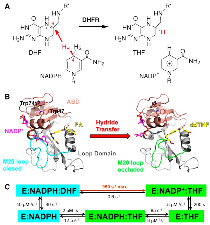
Enzyme motions on a broad range of time scales can play an important role in various intra- and intermolecular events, including substrate binding, catalysis of the chemical conversion, and product release. The relationship between protein motions and catalytic activity is of contemporary interest in enzymology. To understand the factors influencing the rates of enzyme-catalyzed reactions, the dynamics of the protein-solvent-ligand complex must be considered. The current review presents two case studies of enzymes-dihydrofolate reductase (DHFR) and thymidylate synthase (TSase)-and discusses the role of protein motions in their catalyzed reactions. Specifically, we will discuss the utility of kinetic isotope effects (KIEs) and their temperature dependence as tools in probing such phenomena.


Conflict of interest statement

The authors declare no conflict of interest.

Figures

Figure 1
Figure 1
(A) DHFR catalyzes the stereospecific transfer of the pro-R hydride of C4 on NADPH to C6 of protonated N5-DHF, producing THF and the oxidized cofactor NADP+. (B) The active site cleft of DHFR divides the protein into two domains: the adenosine binding domain (ABD, residues 38–88) binds the adenosine moiety of the cofactor NADPH, while the loop domain (~100 residues) is dominated by three loops surrounding the active site. The ternary complex of DHFR with NADP+ (magenta) and folic acid (FA, yellow) mimics the Michaelis complex (structure on the left, PDB code 1RX2). The M20 loop (cyan) closes over the active site to ensure close proximity of the hydride donor (C4 of NADPH) and acceptor (C6 of DHF). The ternary complex of DHFR with NADP+ (magenta) and 5,10-dideazatetrahydrofolic acid (ddTHF, yellow) mimics the product complex (structure on the right, PDB code: 1RX4): here the M20 loop (green) protrudes into the binding site of the nicotinamide ribose moiety of the cofactor to facilitate product release. (C) Under cellular conditions of E. coli (with abundant NADPH concentrations), DHFR cycles through 5 kinetic intermediates, which are colored according to their M20 loop conformations (cyan: closed; green: occluded). The rate constants of each step are from [18]. The maximum (pH-independent) hydride transfer rate (950 s−1) was obtained from non-linear regression of the pH dependence of observed rate constants (pH 5.5–9.0) in stopped-flow experiments [18]. Reprinted from [19] with permission from the American Chemical Society.
Figure 2
Figure 2
Roles of active site and distal residues on the DHFR catalyzed reaction. Left panel: Structure of WT-DHFR (PDB Code 1RX2), with folate in magenta and NADP+ in light blue. A yellow arrow marks the hydride’s path from C4 of the nicotinamide to C6 of the folate, and the residues studied in ref [10] are marked as blue spheres. Right panel: Presentation of the temperature-dependence parameters of intrinsic KIE for WT (black), distal (red), and active site I14 (green) mutants of DHFR, where error bars represent standard deviation. The ordinate is the isotope effect on the activation energy (ΔEa) and the abscissa is the isotope effect on the Arrhenius pre-exponential factor (AH/AT) both of which were determined from a non-linear regression of the intrinsic KIEs to the Arrhenius equation. The yellow block represents the semi-classical range of the Arrhenius pre-exponential factor (0.3–1.7) [49]. Reproduced from ref. [10] with permission from the American Chemical Society.
Figure 3
Figure 3
The KIEs and forward commitment factors (Cf) of light DHFR (l-DHFR; blue) and heavy DHFR (h-DHFR; red) measured from competitive experiments. (A) The observed H/T KIEs (empty symbols) and intrinsic H/T KIEs (filled symbols) are plotted on the logarithmic scale against inverse absolute temperature. The lines are non-linear regression of the intrinsic KIEs to the Arrhenius equation. (B) Cf of h-DHFR is either equal (25–45 °C) to or larger (5–25 °C) than Cf of l-DHFR. Reprinted from ref. [19] with permission from the American Chemical Society.
Scheme 1
Scheme 1
The proposed mechanism for TSase. Adapted from Ref [11] with copyright permission from the American Chemical Society.
Figure 4
Figure 4
(A) The residue-based matrix plots showing correlations of anisotropic B-factor displacements for TSase. The left plot is for WT TSase and the right plot is for Y209W TSase. Segment 2a in the right plot is the phosphate-binding loop with relatively higher B factors. (B) Ribbon diagrams of WT (left) and Y209W (right). The mutated residue, dUMP, catalytic cysteine and cofactor analog (CB3717) are shown as sticks. Reproduced from ref [11] with permission from the American Chemical Society.
Figure 5
Figure 5
(A) Mg2+ binding rigidifies the TSase-(5F-dUMP)-CH2H4fol complex. Significant (greater than 2σ) differences in 15N-1H order parameters (ΔS2 = S2, Mg2+-S2, EDTA) are shown. (B) The significant changes in panel A are highlighted on a ternary complex structure with a color gradient of white to red representing minimal to maximal changes in S2. Residues with no significant change are colored in and the suggested binding site for Mg2+ is shown in green. Reproduced from ref [83] with permission from the American Chemical Society.

References

    1. Wolfenden R. Degrees of Difficulty of Water-Consuming Reactions in the Absence of Enzymes. Chem. Rev. 2006;106:3379–3396. doi: 10.1021/cr050311y. - DOI - PubMed
    1. Benkovic S.J., Hammes-Schiffer S. A Perspective on Enzyme Catalysis. Science. 2003;301:1196–1202. doi: 10.1126/science.1085515. - DOI - PubMed
    1. Jäckel C., Kast P., Hilvert D. Protein Design by Directed Evolution. Annu. Rev. Biochem. Biophys. 2008;37:153–173. doi: 10.1146/annurev.biophys.37.032807.125832. - DOI - PubMed
    1. Fischer E. Influence of configuration on the action of enzymes. Ber. Dtsch. Chem. Ges. 1894;27:2985–2993. doi: 10.1002/cber.18940270364. - DOI
    1. Pauling L. Chemical Achievement and Hope for the Future. Am. Sci. 1948;36:51–58. - PubMed

Publication types

LinkOut - more resources