. 2015 Apr;25(4):598-607.
doi: 10.1101/gr.184184.114. Epub 2015 Jan 15.

A lacZ reporter gene expression atlas for 313 adult KOMP mutant mouse lines

Affiliations

A lacZ reporter gene expression atlas for 313 adult KOMP mutant mouse lines

David B West et al. Genome Res. 2015 Apr.

Abstract

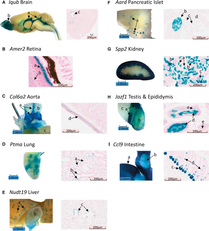
Expression of the bacterial beta-galactosidase reporter gene (lacZ) in the vector used for the Knockout Mouse Project (KOMP) is driven by the endogenous promoter of the target gene. In tissues from KOMP mice, histochemical staining for LacZ enzyme activity can be used to determine gene expression patterns. With this technique, we have produced a comprehensive resource of gene expression using both whole mount (WM) and frozen section (FS) LacZ staining in 313 unique KOMP mutant mouse lines. Of these, ∼ 80% of mutants showed specific staining in one or more tissues, while ∼ 20% showed no specific staining, ∼ 13% had staining in only one tissue, and ∼ 25% had staining in >6 tissues. The highest frequency of specific staining occurred in the brain (∼ 50%), male gonads (42%), and kidney (39%). The WM method was useful for rapidly identifying whole organ and some substructure staining, while the FS method often revealed substructure and cellular staining specificity. Both staining methods had >90% repeatability in biological replicates. Nonspecific LacZ staining occurs in some tissues due to the presence of bacteria or endogenous enzyme activity. However, this can be effectively distinguished from reporter gene activity by the combination of the WM and FS methods. After careful annotation, LacZ staining patterns in a high percentage of mutants revealed a unique structure-function not previously reported for many of these genes. The validation of methods for LacZ staining, annotation, and expression analysis reported here provides unique insights into the function of genes for which little is currently known.


Figures

Figure 1.
Figure 1.
Patterns of LacZ staining in whole mount (WM, black) and frozen section (FS, gray) preparations. Male and female data are combined for this analysis. For calculating the percentage of organs/tissues stained, brain is counted as one organ whether one or six different brain regions stain. Overall, similar patterns and percentages of tissues stained using the two methods.
Figure 2.
Figure 2.
Frequency of nonspecific staining in wild-type (WT) control C57BL/6N male and female mice. Whole mount (WM), and frozen section (FS) nonspecific staining percentages are indicated in blue and red, respectively. Explanations of differences between WM and FS preparations for nonspecific staining for specific tissues are as follows: (1) bacteria on epithelium of tongue and esophagus in FS preparations; (2) luminal gut bacteria present in intact WM preparations; (3) prostate adjacent to preputial gland and inability to distinguish between preputial gland nonspecific staining and specific prostate gland staining in WM preparations; and (4) likely differences in staining conditions allowing the detection of endogenous galactoside activity in either WM or FS preparations, but not both. Nonspecific staining is particularly problematic in the GI tract and in the male sex organs.
Figure 3.
Figure 3.
(A) Wtip olfactory bulb, cerebral, and cerebellar blood vessels stain for LacZ in lateral brain whole mount (WM, left), and frozen section (FS, right) reveals staining of a branching cerebral blood vessel. In the Wtip brain, only blood vessels stain for LacZ. (B) Prr15l staining of lung in WM (left) and FS (right). In WM, the major airways clearly stain, but there is also a diffuse staining of the parenchymal tissue. In the FS image, the intense staining of the bronchiole epithelium (a), and the weaker punctate pattern of staining in the walls of the alveoli (b) are indicated. (C, left) Dram1 staining of mesenteric adipose tissue by WM showing diffuse uniform staining (a), while the ileum does not stain (b). (Right) FS of Dram1 mesenteric adipose tissue shows staining is restricted to the stromal, intercellular space (c). (D, left) Zfp12 WM staining of fundus (a), body (b), and antrum (c) of the stomach. WM staining of the pyloris and duodenum (d) and weak staining of the esophagus (e) are similar to that found in WT mice. (Right) FS indicates that the majority of Zfp12 LacZ staining is in the smooth muscle layer (f), although there is weak, scattered staining in the glandular epithelium (g). (E, left) Sec23a WM staining of cartilaginous ribs (a), intersternal plate cartilage (b), blood vessels (c), and a weak striated pattern of staining of the chest muscle (d). (Right) FS of striated Sec23a muscle indicates staining of connective tissue and/or blood vessels (e,f) between muscle fibers.
Figure 4.
Figure 4.
(A) Iqub whole mount (WM, left) mid-sagittal section showing staining of ventricular ependyma throughout the brain including the lateral and fourth ventricles (a), as well as staining of the glomeruli in the olfactory bulb (b), and coronal frozen section (FS, right) staining of forebrain illustrating ependymal staining of the lateral ventricle (c). (B) Amer2 FS retina with intense staining of the photoreceptor cell layer (a) and diffuse scattered staining of the inner nuclear layer (b). (C, left) Col6a2 aorta staining in WM showing distinct staining of the aortic arch and the carotid arteries (a) with no staining of white adipose tissue (b) or the left ventricle (c). (Right) FS of the aorta shows smooth muscle only staining for LacZ (d). (D, left) Ptma WM lung staining showing a variegated staining pattern. (Right) This uneven, variegated pattern of staining is also found in FS with individual cells staining in the parenchyma (a), as well as nonciliated cells in the bronchial epithelium (b). (E, left) Nudt19 liver staining is shown for WM, indicating that the bile ducts (a) and gall bladder (b) stain for LacZ. (Right) FS staining shows that epithelial cells in the ducts are expressing the lacZ reporter. (F, left) Aard pancreas staining by the WM method shows a distributed punctate staining pattern suggestive of islets. (Right) Aard FS confirms the inference of islet staining from the WM studies and further shows that a subset of cells stains in the islet (b), while ducts (c) and exocrine tissue (d) do not stain in Aard. (G, left) Spp2 kidney staining by WM shows intense staining of the tubules in the cortex only, with no staining in the medulla or hilus. (Right) Spp2 FS shows renal cortical staining in tubules (a), Bowman's capsule (b), and some cells/membranes in the interior of Bowman's capsule (c). (H, left) Jazf1 WM testis staining shows the characteristic WT nonspecific pattern of staining in the epididymis (a) with a 2-mm-wide band of minimal staining separating caput LacZ and additional LacZ staining in the body of the epididymis. Although faint, there is above-background WM staining in the seminiferous tubules (b) within the intact tunica, while the associated adipose tissue (c) does not stain. (Right) FS staining of Jazf1 shows intense staining of seminiferous tubule lumen (d), suggesting that immature sperm express the lacZ reporter. Some, but not all spermatogonia also appear to stain in the FS (e). (I, left) Ccl9 intestinal staining by WM shows a uniform punctate pattern of staining in the wall of the small intestine (a). The vasculature is clearly demarked on the surface of the intestine since it does not stain. Peyer's patches (b) also do not stain, except for a small band of staining encircling each dome in the patch (b). (Right) FS staining of Ccl9 shows staining primarily in the Crypts of Lieberkuhn (c), but there are some cells in the microvilli also staining for LacZ (d).

References

    1. Adams AC, Gale NW. 2006. High resolution gene expression analysis in mice using genetically inserted reporter genes. In Mammalian and avian transgenesis: new approaches (principles and practice) (ed. Pease S, Lois C), pp. 131–161 Springer-Verlag, Berlin.
    1. Bradley A, Anastassiadis K, Ayadi A, Battey JF, Bell C, Birling MC, Bottomley J, Brown SD, Bürger A, Bult CJ, et al.2012. The mammalian gene function resource: the International Knockout Mouse Consortium. Mamm Genome 23: 580–586. - PMC - PubMed
    1. Brown SD, Moore MW. 2012. The International Mouse Phenotyping Consortium: past and future perspectives on mouse phenotyping. Mamm Genome 23: 632–640. - PMC - PubMed
    1. Casadaban MJ, Chou J, Cohen SN. 1980. In vitro gene fusions that join an enzymatically active β-galactosidase segment to amino-terminal fragments of exogenous proteins: Escherichia coli plasmid vectors for the detection and cloning of translational initiation signals. J Bacteriol 143: 971–980. - PMC - PubMed
    1. Gates H, Mallon AM, Brown SD; EUMODIC Consortium. 2011. High-throughput mouse phenotyping. Methods 53: 394–404. - PubMed

Publication types