. 2015 Feb 17;87(4):2273-81.
doi: 10.1021/ac504118y. Epub 2015 Jan 27.

Avoiding misannotation of in-source fragmentation products as cellular metabolites in liquid chromatography-mass spectrometry-based metabolomics

Affiliations

Avoiding misannotation of in-source fragmentation products as cellular metabolites in liquid chromatography-mass spectrometry-based metabolomics

Yi-Fan Xu et al. Anal Chem. .

Abstract

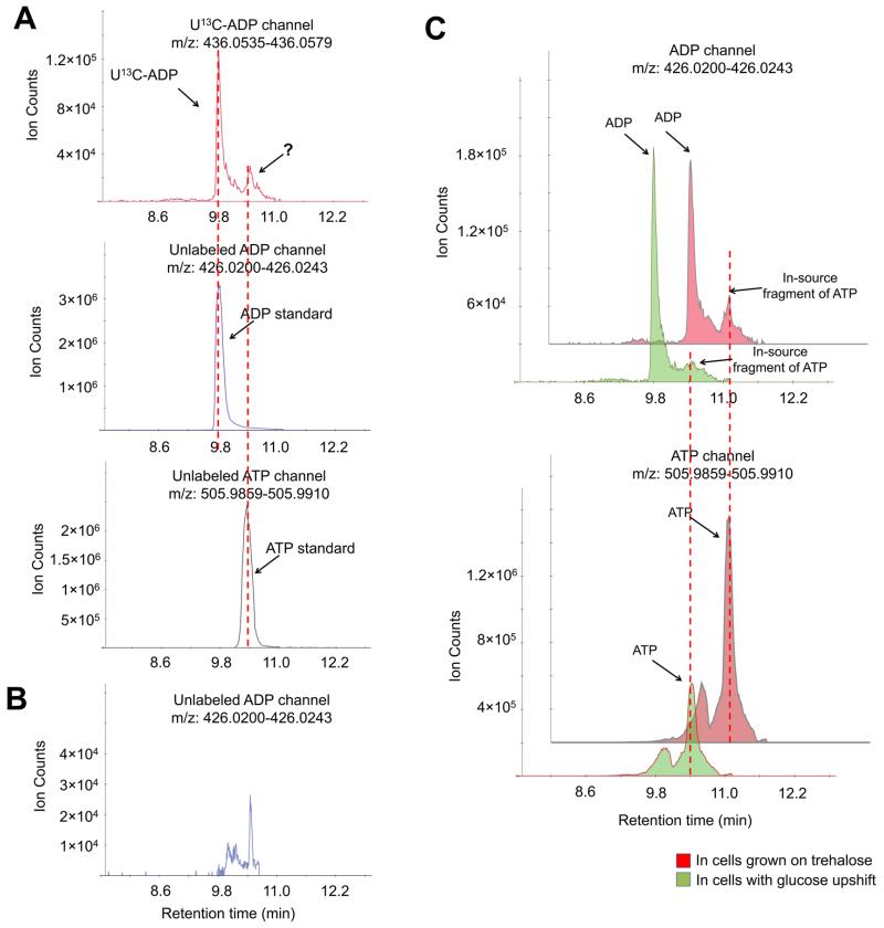
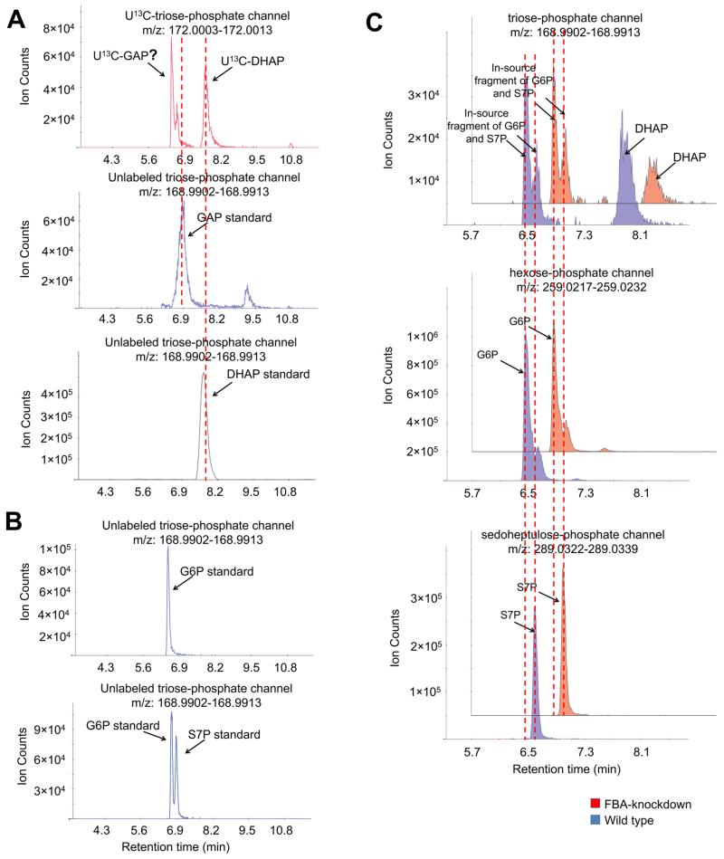
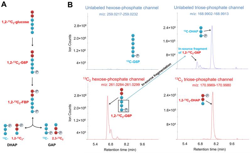
Liquid chromatography-mass spectrometry (LC-MS) technology allows for rapid quantitation of cellular metabolites, with metabolites identified by mass spectrometry and chromatographic retention time. Recently, with the development of rapid scanning high-resolution high accuracy mass spectrometers and the desire for high throughput screening, minimal or no chromatographic separation has become increasingly popular. When analyzing complex cellular extracts, however, the lack of chromatographic separation could potentially result in misannotation of structurally related metabolites. Here, we show that, even using electrospray ionization, a soft ionization method, in-source fragmentation generates unwanted byproducts of identical mass to common metabolites. For example, nucleotide-triphosphates generate nucleotide-diphosphates, and hexose-phosphates generate triose-phosphates. We evaluated yeast intracellular metabolite extracts and found more than 20 cases of in-source fragments that mimic common metabolites. Accordingly, chromatographic separation is required for accurate quantitation of many common cellular metabolites.


Figures

Figure 1
Figure 1
Correct quantitation of ADP requires chromatographic separation. (A) The negative ionization mode-extracted ion chromatogram for U13C-ADP (+10, upper panel) and unlabeled ADP (lower panel). Yeast cells were grown in U13C-glucose, and metabolome was extracted with quenching solution spiked with unlabeled ADP. (B) The negative ionization mode-extracted ion chromatogram for the ADP channel in the ATP standard. The retention time of the “ADP” peak matched the retention time of the ATP standard, indicating that such a peak is an in-source fragment. (C) The negative ionization mode-extracted ion chromatogram for ADP and ATP channels in yeast cells grown on trehalose and 5 min after switching to glucose. Method A has been used throughout this figure.
Figure 2
Figure 2
Glyceraldehyde-3-phosphate peak is masked by in-source fragments of glucose-6-phosphate and sedoheptulose-7-phosphate. (A) The negative ionization mode-extracted ion chromatogram for U13C-triose-phosphate (+3, upper panel) and unlabeled triose-phosphate (lower panel). Yeast cells were grown in U13C-glucose, and metabolome was extracted with quenching solution spiked with unlabeled glyceraldehyde-3-phosphate (GAP). (B) The negative ionization mode-extracted ion chromatogram for “triose-phosphate” peaks in the glucose-6-phosphate and sedoheptulose-7-phosphate standards. Glucose-6-phosphate (upper panel) and a mixture of glucose-6-phosphate and sedoheptulose-7-phosphate standards (lower panel) were analyzed by LC-MS. The retention time of the “triose-phosphate” peaks matched glucose-6-phosphate and sedoheptulose-7-phosphate standards, indicating that they are in-source fragments. (C) The negative ionization mode-extracted ion chromatogram for triose-phosphate, glucose-6-phosphate, and sedoheptulose-7-phosphate channels in wild type and FBA knockdown yeast cells. Wild type and FBA knockdown yeast cells were grown to exponential phase, and their metabolome was extracted and analyzed by LC-MS. Method A has been used throughout this figure.
Figure 3
Figure 3
Confirmation via isotope labeling that the putative glyceraldehyde-3-phosphate peak is from in-source fragmentation. (A) Schematic of upper glycolytic intermediates in yeast cells grown on 1,2-13C2-glucose. (B). The negative ionization mode-extracted ion chromatogram for unlabeled and 13C2 hexose-phosphate and triose-phosphate channels. Dihydroxyacetone-phosphate (DHAP) shows up in both unlabeled and 13C2 channels, but the leftmost “triose-phosphate” peak only shows up in the unlabeled channel, confirming that this peak is the in-source fragmentation of the last three carbons of glucose-6-phosphate. Method A has been used throughout this figure.

References

    1. Kuehnbaum NL, Britz-McKibbin P. Chem. Rev. 2013;113:2437–2468. - PubMed
    1. Milne SB, Mathews TP, Myers DS, Ivanova PT, Brown HA. Biochemistry. 2013;52:3829–3840. - PMC - PubMed
    1. Theodoridis G, Gika HG, Wilson ID. Mass Spectrom. Rev. 2011;30:884–906. - PubMed
    1. Zhou B, Xiao JF, Tuli L, Ressom HW. Mol. BioSyst. 2012;8:470–481. - PMC - PubMed
    1. Kueger S, Steinhauser D, Willmitzer L, Giavalisco P. Plant J. Cell Mol. Biol. 2012;70:39–50. - PubMed

Publication types