

Review
doi: 10.1038/nature14188.
From circuits to behaviour in the amygdala
Affiliations
- PMID: 25592533
- PMCID: PMC4565157
- DOI: 10.1038/nature14188
Item in Clipboard
Review
From circuits to behaviour in the amygdala
Nature.
.
Abstract
The amygdala has long been associated with emotion and motivation, playing an essential part in processing both fearful and rewarding environmental stimuli. How can a single structure be crucial for such different functions? With recent technological advances that allow for causal investigations of specific neural circuit elements, we can now begin to map the complex anatomical connections of the amygdala onto behavioural function. Understanding how the amygdala contributes to a wide array of behaviours requires the study of distinct amygdala circuits.
Figures
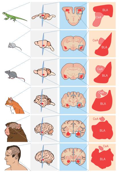
Primary amygdalar nuclei and basic circuit connections and function are conserved across species. An enlarged image of the basolateral complex of the amygdala (BLA) and central nucleus of the amygdala (CeA) or analogues are shown next to a coronal section from the brains of a lizard, mouse, rat, cat, monkey and human.
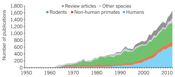
Publications on the amygdala indexed on PubMed between 1950 and 2013 demonstrate the growing interest in amygdala research.
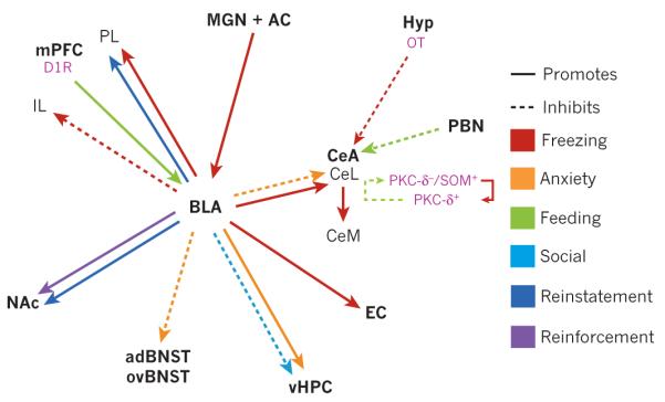
Projection-specific effects as shown by optogenetic or pharmacogenetic manipulation. The solid or dotted lines indicate the promotion or inhibition of certain behaviours. The basolateral complex of the amygdala (BLA) encompasses the lateral and basal nuclei. Specific cell types are shown in pink. For simplicity, projections that are anatomically or electrophysiologically defined but have not been shown to have a causal relationship with behaviour are omitted. This is a selective picture of projections that have been directly manipulated, and is not meant to signify their importance over other anatomical connections. The actual connectivity of the amygdala with other brain regions is considerably more complex. AC, auditory cortex; adBNST, anterodorsal bed nucleus of the stria terminalis; CeA, central nucleus of the amygdala; CeL, lateral CeA; CeM, medial CeA; D1R, dopamine 1 receptor; EC, entorhinal cortex; Hyp, hypothalamus; IL, infralimbic; MGN, medial geniculate nucleus; mPFC, medial prefrontal cortex; NAc, nucleus accumbens; OT, oxytocin; ovBNST, oval nucleus of the BNST; PBN, parabrachial nucleus; PKC, protein kinase C; PL, prelimbic; SOM, somatostatin; vHPC, ventral hippocampus.
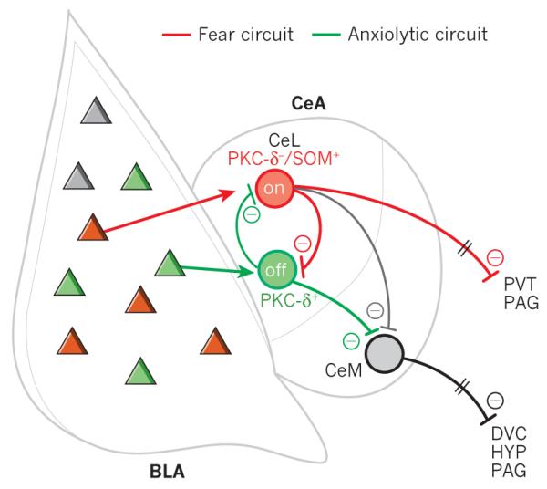
New findings in the amgydala have updated our understanding of these microcircuits. Different populations of basolateral complex of the amygdala (BLA) neurons are proposed to activate distinct populations of lateral central nucleus of the amygdala (CeL) neurons to either promote fear or reduce anxiety. CeM, medial central nucleus of the amygdala; DVC, dorsal vagal complex; PAG, periaqueductal grey; PKC, protein kinase C; PVT, paraventricular nucleus of the thalamus; HYP, hypothalamus; SOM, somatostatin.
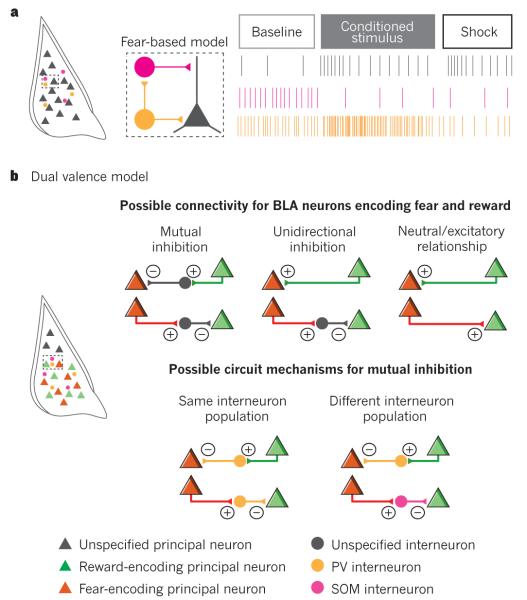
a, Model of how interneurons expressing parvalbumin (PV) and somatostatin (SOM) interact with principal neurons to mediate fear conditioning. b, The heterogeneity in PV interneuron responses is consistent with the diverse functionality of BLA principal neurons and raises the question of how BLA principal neurons may interact locally. Depicted are simplified scenarios for these interactions.
References
-
- McDonald AJ. Cortical pathways to the mammalian amygdala. Prog. Neurobiol. 1998;55:257–332. - PubMed
-
- Johnston JB. Further contributions to the study of the evolution of the forebrain. J. Comp. Neurol. 1923;35:337–481.
-
- Kappers CUA, Huber GC, Crosby EC. Including Man. Macmillan; 1936. The Comparative Anatomy of the Nervous System of Vertebrates.
-
- Lanuza E, Belekhova M, Martínez-Marcos A, Font C, Martínez-García F. Identification of the reptilian basolateral amygdala: an anatomical investigation of the afferents to the posterior dorsal ventricular ridge of the lizard Podarcis hispanica. Eur. J. Neurosci. 1998;10:3517–3534. - PubMed
Publication types
MeSH terms
Grants and funding
LinkOut - more resources
Full Text Sources
Other Literature Sources
Medical
