

Manipulating cell fate in the cochlea: a feasible therapy for hearing loss
- PMID: 25593106
- PMCID: PMC4352405
- DOI: 10.1016/j.tins.2014.12.004
Manipulating cell fate in the cochlea: a feasible therapy for hearing loss
Abstract
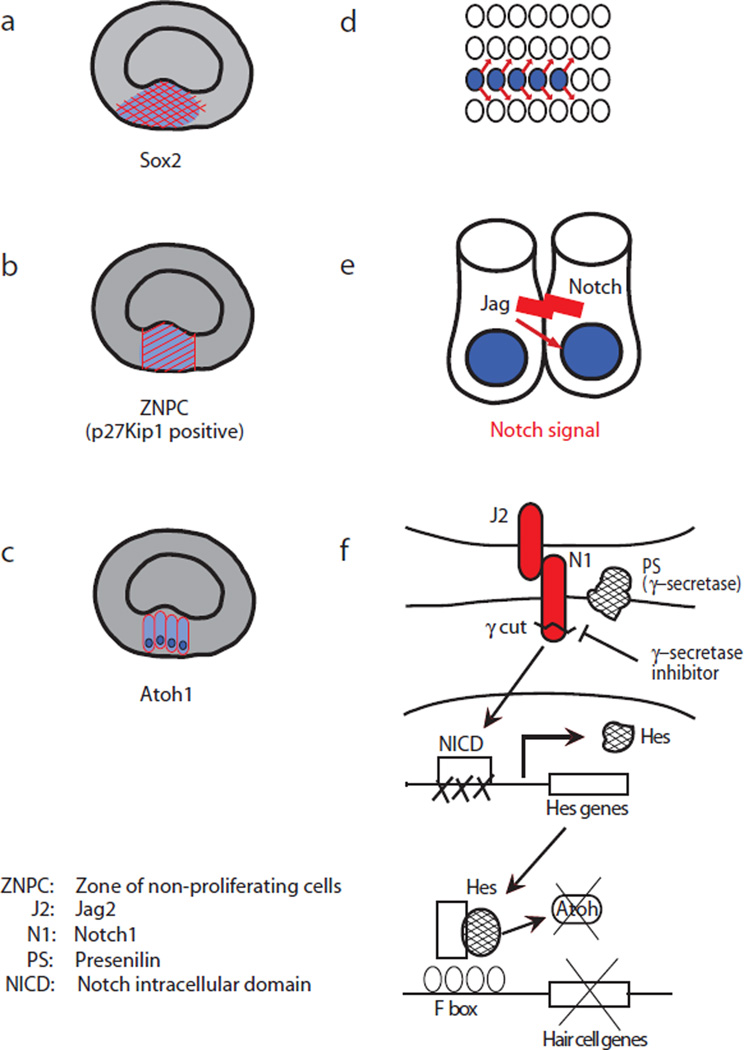
Mammalian auditory hair cells do not spontaneously regenerate, unlike hair cells in lower vertebrates, including fish and birds. In mammals, hearing loss due to the loss of hair cells is permanent and intractable. Recent studies in the mouse have demonstrated spontaneous hair cell regeneration during a short postnatal period, but this regenerative capacity is lost in the adult cochlea. Reduced regeneration coincides with a transition that results in a decreased pool of progenitor cells in the cochlear sensory epithelium. Here, we review the signaling cascades involved in hair cell formation and morphogenesis of the organ of Corti in developing mammals, the changing status of progenitor cells in the cochlea, and the regeneration of auditory hair cells in adult mammals.
Keywords: cell replacement; hair cells; hearing loss; sensory systems.
Copyright © 2014 Elsevier Ltd. All rights reserved.
Figures

Similar articles
-
Hearing restoration through hair cell regeneration: A review of recent advancements and current limitations.Hear Res. 2025 Jun;461:109256. doi: 10.1016/j.heares.2025.109256. Epub 2025 Mar 22. Hear Res. 2025. PMID: 40157114 Review.
-
Recent advances in cochlear hair cell regeneration-A promising opportunity for the treatment of age-related hearing loss.Ageing Res Rev. 2017 Jul;36:149-155. doi: 10.1016/j.arr.2017.04.002. Epub 2017 Apr 13. Ageing Res Rev. 2017. PMID: 28414155 Review.
-
Role of Wnt and Notch signaling in regulating hair cell regeneration in the cochlea.Front Med. 2016 Sep;10(3):237-49. doi: 10.1007/s11684-016-0464-9. Epub 2016 Sep 7. Front Med. 2016. PMID: 27527363 Review.
-
Transdifferentiation and its applicability for inner ear therapy.Hear Res. 2007 May;227(1-2):41-7. doi: 10.1016/j.heares.2006.08.015. Epub 2006 Oct 27. Hear Res. 2007. PMID: 17070000 Review.
-
Postnatal development of the hamster cochlea. I. Growth of hair cells and the organ of Corti.J Comp Neurol. 1994 Feb 1;340(1):87-97. doi: 10.1002/cne.903400107. J Comp Neurol. 1994. PMID: 8176004
Cited by
-
Base editing: advances and therapeutic opportunities.Nat Rev Drug Discov. 2020 Dec;19(12):839-859. doi: 10.1038/s41573-020-0084-6. Epub 2020 Oct 19. Nat Rev Drug Discov. 2020. PMID: 33077937 Free PMC article. Review.
-
Gene therapy: an emerging therapy for hair cells regeneration in the cochlea.Front Neurosci. 2023 May 3;17:1177791. doi: 10.3389/fnins.2023.1177791. eCollection 2023. Front Neurosci. 2023. PMID: 37207182 Free PMC article. Review.
-
Additive reductions in zebrafish PRPS1 activity result in a spectrum of deficiencies modeling several human PRPS1-associated diseases.Sci Rep. 2016 Jul 18;6:29946. doi: 10.1038/srep29946. Sci Rep. 2016. PMID: 27425195 Free PMC article.
-
Inner Ear Organoid as a Preclinical Model of Hearing Regeneration: Progress and Applications.Stem Cell Rev Rep. 2025 Jul 25. doi: 10.1007/s12015-025-10941-5. Online ahead of print. Stem Cell Rev Rep. 2025. PMID: 40711673 Review.
-
Recent advancements in understanding the role of epigenetics in the auditory system.Gene. 2020 Nov 30;761:144996. doi: 10.1016/j.gene.2020.144996. Epub 2020 Jul 29. Gene. 2020. PMID: 32738421 Free PMC article. Review.
References
-
- Bermingham NA, et al. Math1: an essential gene for the generation of inner ear hair cells. Science. 1999;284(5421):1837–1841. - PubMed
-
- Lanford PJ, et al. Notch signalling pathway mediates hair cell development in mammalian cochlea. Nat Genet. 1999;21(3):289–292. - PubMed
-
- Woods C, Montcouquiol M, Kelley MW. Math1 regulates development of the sensory epithelium in the mammalian cochlea. Nat Neurosci. 2004;7(12):1310–1318. - PubMed
-
- Hayashi T, Cunningham D, Bermingham-McDonogh O. Loss of Fgfr3 leads to excess hair cell development in the mouse organ of Corti. Dev Dyn. 2007;236(2):525–533. - PubMed
Publication types
MeSH terms
Grants and funding
LinkOut - more resources
Full Text Sources
Other Literature Sources
