. 2015 Feb;4(2):165-77.
doi: 10.5966/sctm.2014-0179. Epub 2015 Jan 15.

Defined culture of human embryonic stem cells and xeno-free derivation of retinal pigmented epithelial cells on a novel, synthetic substrate

Affiliations

Defined culture of human embryonic stem cells and xeno-free derivation of retinal pigmented epithelial cells on a novel, synthetic substrate

Britney O Pennington et al. Stem Cells Transl Med. 2015 Feb.

Abstract

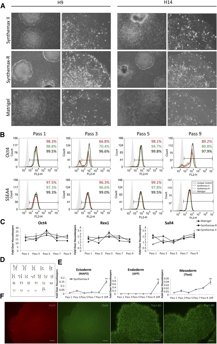
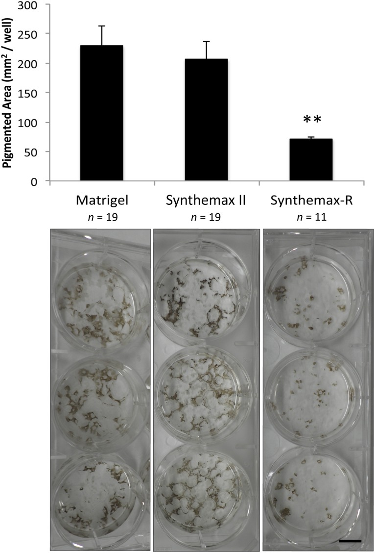
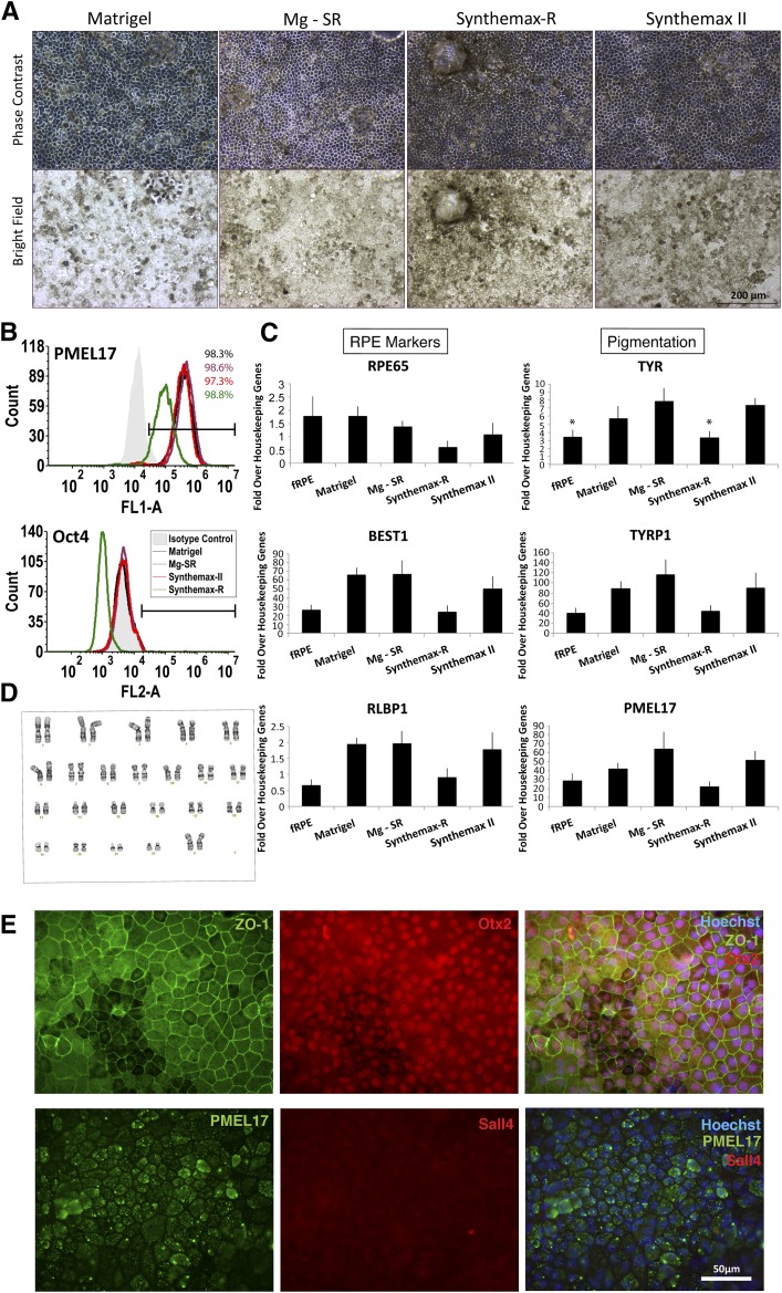
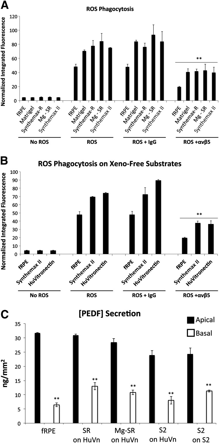
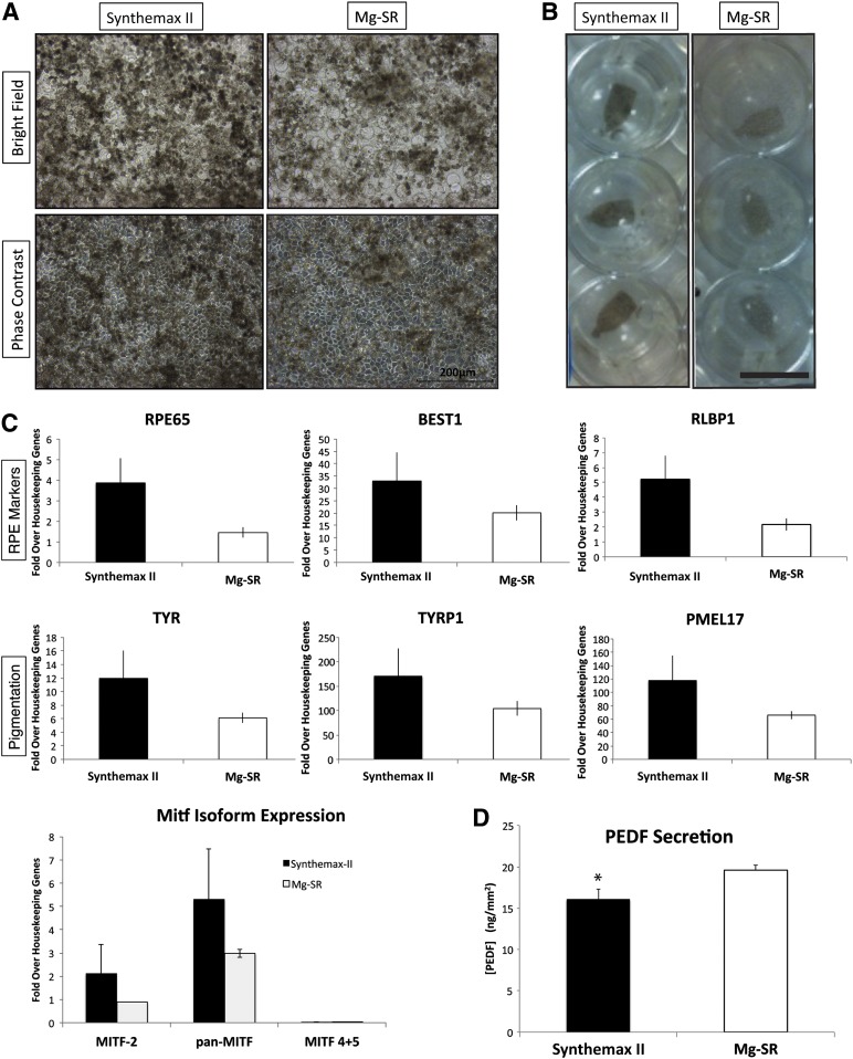
Age-related macular degeneration (AMD), a leading cause of blindness, is characterized by the death of the retinal pigmented epithelium (RPE), which is a monolayer posterior to the retina that supports the photoreceptors. Human embryonic stem cells (hESCs) can generate an unlimited source of RPE for cellular therapies, and clinical trials have been initiated. However, protocols for RPE derivation using defined conditions free of nonhuman derivatives (xeno-free) are preferred for clinical translation. This avoids exposing AMD patients to animal-derived products, which could incite an immune response. In this study, we investigated the maintenance of hESCs and their differentiation into RPE using Synthemax II-SC, which is a novel, synthetic animal-derived component-free, RGD peptide-containing copolymer compliant with good manufacturing practices designed for xeno-free stem cell culture. Cells on Synthemax II-SC were compared with cultures grown with xenogeneic and xeno-free control substrates. This report demonstrates that Synthemax II-SC supports long-term culture of H9 and H14 hESC lines and permits efficient differentiation of hESCs into functional RPE. Expression of RPE-specific markers was assessed by flow cytometry, quantitative polymerase chain reaction, and immunocytochemistry, and RPE function was determined by phagocytosis of rod outer segments and secretion of pigment epithelium-derived factor. Both hESCs and hESC-RPE maintained normal karyotypes after long-term culture on Synthemax II-SC. Furthermore, RPE generated on Synthemax II-SC are functional when seeded onto parylene-C scaffolds designed for clinical use. These experiments suggest that Synthemax II-SC is a suitable, defined substrate for hESC culture and the xeno-free derivation of RPE for cellular therapies.

Keywords: Age-related macular degeneration; Human embryonic stem cells; Parylene-C; Retinal pigmented epithelium; Synthemax II-SC substrate.


Figures

Figure 1.
Figure 1.
Characterization of human embryonic stem cells (hESCs) cultured on Synthemax II-SC, Synthemax-R, and Matrigel. (A): Representative phase-contrast micrographs of H9 and H14 hESC colony morphology after nine passages on the indicated substrate. Scale bars = 200 μm. (B): Representative flow cytometry histograms for pluripotency markers Oct4 and SSEA4 in H9 cultures grown on the indicated substrate at passages 1, 3, 5, and 9. (C): Relative expression of pluripotency genes in H9 cultures grown on Synthemax II-SC, Synthemax-R, and Matrigel at passages 1, 3, 5, and 9 as detected by quantitative polymerase chain reaction. (D): Normal karyotype of H9 hESCs maintained on Synthemax II-SC for 23 passages. (E): Relative gene expression of germ layer markers is significantly higher in H9 hESCs differentiated on Synthemax II-SC for 10 days compared with hESCs at earlier passages. ∗∗, p < 0.01, t test. (F): H9 hESCs on Synthemax II-SC stained for nuclear pluripotency transcription factors Oct4 and Sall4 and surface markers Tra-1-81 and Tra-1-60. Scale bars = 80 μm. Abbreviation: Diff, differentiation.
Figure 2.
Figure 2.
Spontaneous differentiation of retinal pigmented epithelia generated from human embryonic stem cells (hESC-RPE) on Matrigel and Synthemax II-SC yields significantly more pigmented area than hESC-RPE on Synthemax-R. Top: Pigmented area in individual wells was calculated with ImageJ software after 115 days of differentiation. Error bars denote standard deviation. ∗∗, p < .01, t test. Bottom: Three representative wells from a six-well plate of differentiated hESC-RPE seeded on each substrate are shown. Scale bar = 1 cm.
Figure 3.
Figure 3.
Characterization of RPE generated from human embryonic stem cells (hESC-RPE) derived on Synthemax II-SC. (A): Phase-contrast and bright-field images show the typical pigmentation and cobblestone morphology of H9 hESC-RPE cultured on different surfaces are shown. Scale bar = 200 μm. (B): Representative flow cytometry histograms are shown for the premelanosome pigmentation marker PMEL17 and the pluripotency marker Oct4 (not detected) in H9-RPE at passage 2 day 28 on the indicated substrate. (C): Expression of RPE marker genes in H9 hESC-RPE on the indicated substrates. fRPE served as a positive control (n = 3). ∗, p < .05, t test. (D): Normal karyotype of H9 hESC-RPE after spontaneous differentiation, enrichment, and three passages on Synthemax II-SC. (E): Epifluorescent images are shown of H9 hESC-RPE derived on Synthemax II-SC stained for the tight junction marker, ZO-1, RPE markers Otx2 and PMEL17, and the pluripotency marker Sall4 (not detected). Nuclei were detected using Hoechst (blue, merged right panels). Scale bar = 50 μm. Abbreviations: fRPE, fetal retinal pigmented epithelium; Mg-SR, condition of culturing and differentiating hESCs on Matrigel and then enriching and propagating the hESC-RPE on Synthemax-R; RPE, retinal pigmented epithelium.
Figure 4.
Figure 4.
Functional characterization of H9 RPE generated from human embryonic stem cells (hESC-RPE) derived on Synthemax II-SC. (A): Phagocytosis of ROS by H9 hESC-RPE derived on Matrigel, Synthemax-R, Synthemax II-SC, or Mg-SR. A function-blocking antibody against αvβ5 integrin, which is necessary for phagocytosis by RPE, significantly decreased internalization of ROS in all hESC-RPE when compared with the IgG isotype control. ∗∗, p < .01, t test. fRPE served as a positive control. (B): Phagocytosis of ROS by H9 hESC-RPE derived on Synthemax II-SC after seeding on the defined substrates Synthemax II-SC and human vitronectin. Phagocytic activity was significantly decreased with the function-blocking αvβ5 antibody when compared with the IgG isotype control. ∗∗, p < .01, t test. (C): All conditions secrete PEDF significantly more on the apical surface compared with the basal side when seeded on surfaces coated with human vitronectin or Synthemax II-SC. ∗∗, p < .01, t test. hESC-RPE derived on Synthemax II-SC secrete less apical PEDF compared with fRPE and cells derived on Synthemax-R. Abbreviations: fRPE, fetal retinal pigmented epithelium; HuVn, human vitronectin; ROS, rod outer segment; S2, hESC-RPE derived on Synthemax II-SC; Mg-SR, hESCs grown and differentiated on Matrigel and then enriched as hESC-RPE on Synthemax-R; PEDF, pigment epithelium-derived factor; SR, hESC-RPE derived on Synthemax-R.
Figure 5.
Figure 5.
Characterization of H9 RPE generated from human embryonic stem cells (hESC-RPE) derived on Synthemax II-SC versus Mg-SR seeded onto parylene-C scaffolds. (A): Bright-field and phase-contrast images of H9 hESC-RPE cells grown for 30 days on parylene-C scaffolds. hESC-RPE were derived on Synthemax II-SC or by the Mg-SR method. Ultrathin regions of the membrane appear as an array of circles in the micrographs. Scale bar = 200 μm. (B): Bright-field images of three representative parylene-C scaffolds seeded with hESC-RPE in a 24-well plate (n = 8). Scale bar = 1 cm. Note darker pigmentation in the RPE derived on Synthemax II-SC. (C): Expression of RPE and pigmentation markers after 30 days on parylene-C scaffolds as detected by quantitative polymerase chain reaction (n = 3). (D): Quantification of PEDF protein secreted by hESC-RPE derived on Synthemax II-SC or by the Mg-SR method after 30 days on parylene-C scaffolds (n = 6; error is SEM). ∗, p < .05, t test. Abbreviations: Mg-SR, hESCs grown and differentiated on Matrigel and then enriched as hESC-RPE on Synthemax-R; PEDF, pigment epithelium-derived factor; RPE, retinal pigmented epithelium.

Similar articles

Cited by

References

    1. Rein DB, Zhang P, Wirth KE, et al. The economic burden of major adult visual disorders in the United States. Arch Ophthalmol. 2006;124:1754–1760. - PubMed
    1. Wong WL, Su X, Li X, et al. Global prevalence of age-related macular degeneration and disease burden projection for 2020 and 2040: A systematic review and meta-analysis. Lancet Glob Health. 2014;2:e106–e116. - PubMed
    1. Khandhadia S, Cherry J, Lotery AJ. Age-related macular degeneration. Adv Exp Med Biol. 2012;724:15–36. - PubMed
    1. Young RW, Bok D. Participation of the retinal pigment epithelium in the rod outer segment renewal process. J Cell Biol. 1969;42:392–403. - PMC - PubMed
    1. Miller SS, Edelman JL. Active ion transport pathways in the bovine retinal pigment epithelium. J Physiol. 1990;424:283–300. - PMC - PubMed

Publication types

MeSH terms