Review
. 2015 Jan 15;29(2):109-22.
doi: 10.1101/gad.255554.114.

From equator to pole: splitting chromosomes in mitosis and meiosis

Affiliations
Review

From equator to pole: splitting chromosomes in mitosis and meiosis

Eris Duro et al. Genes Dev. .

Abstract

During eukaryotic cell division, chromosomes must be precisely partitioned to daughter cells. This relies on a mechanism to move chromosomes in defined directions within the parental cell. While sister chromatids are segregated from one another in mitosis and meiosis II, specific adaptations enable the segregation of homologous chromosomes during meiosis I to reduce ploidy for gamete production. Many of the factors that drive these directed chromosome movements are known, and their molecular mechanism has started to be uncovered. Here we review the mechanisms of eukaryotic chromosome segregation, with a particular emphasis on the modifications that ensure the segregation of homologous chromosomes during meiosis I.

Keywords: kinetochore; meiosis; microtubules; mitosis.


Figures

Figure 1.
Figure 1.
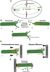
Microtubules drive chromosome motion. (A) The different types of microtubules (light green) nucleated by the centrosome (dark green): Astral microtubules project into the cell cortex, kinetochore microtubules connect the poles to the chromosomes to be segregated, and interpolar microtubules interdigitate to provide structural rigidity. (B) The coexistence of assembly and disassembly at the plus end of microtubules is described as dynamic instability. Microtubules grow via the addition of GTP-bound α-tubulin and β-tubulin dimers. (C) The energy released from GTP hydrolysis during microtubule assembly is stored in the polymer lattice via the geometrical constraint imposed by the bend. (D) Two different models of how microtubule depolymerization can provide the energy for directional motion of chromosomes. (Left panel) In the conformational wave model, as the disassembling protofilaments curve outward, a “sliding collar” (often posited to be a ring; see the text) is driven toward the minus end. (Right panel) In the biased diffusion model, a binding free-energy gradient ensures biased direction. (E) Addition of tubulin subunits to kinetochore-bound microtubule plus ends counteracts the loss of tubulin subunits from the minus ends, thus creating a constant poleward flow of tubulin subunits. This poleward flux is thought to contribute to correct microtubule attachment and chromosome motion.
Figure 2.
Figure 2.
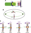
Kinetochore–microtubule interactions. (A) Diagram of the organization of the kinetochore. The inner kinetochore (purple) assembles on the centromeres of chromosomes (gray). The outer kinetochore (SPC105 [light blue] NDC80 [yellow], and DASH [blue ring]) forms the microtubule-binding interface. The central kinetochore (red) links the inner and outer subcomplexes of the kinetochore. The DASH complex ring is yeast-specific but is thought to be functionally analogous to the Ska1 complex in higher organisms. (B) Microtubules are first captured laterally by the kinetochore (red circles). These are then converted into stronger and more processive end-on attachments. The black triangles indicate kinetochore orientation. (C) Tension is generated when the pulling force of the microtubule is counteracted by cohesion (blue rings) holding sister chromatids together (see the text). Sister kinetochores are capable of capturing microtubules emanating from either spindle pole. Attachments that do not generate even tension allow Aurora B kinase (yellow) to sever kinetochore–microtubule attachments. Amphitelic attachments generate equal tension across sister kinetochores, thus removing Aurora B substrates from its reach.
Figure 3.
Figure 3.
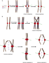
The chromosome segregation program in mitosis (A) and meiosis (B). During DNA replication, cohesin rings (blue) topologically entrap sister DNA molecules to give rise to sister chromatid cohesion. (A) In mitotic metaphase, sister kinetochores (red circles) are bioriented (black triangles): They attach to microtubules (green) emanating from opposite spindle poles. In anaphase, cohesin is cleaved, allowing sister chromatids to separate. (B) In meiotic prophase I, homologous recombination (HR) allows homologous chromosomes to be physically linked via chiasmata. In meiotic metaphase I, sister kinetochores are thought to fuse so as to present as a single microtubule-binding interface. In anaphase I, centromere-distal cohesin is cleaved, allowing homologous chromosomes to separate. Centromere cohesin, however, is protected. In meiosis II, much like in mitosis, sister kinetochores biorient in metaphase II, and the cleavage of centromere cohesin in anaphase II allows sister chromatids to segregate.
Figure 4.
Figure 4.
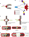
Mono-orientation of sister kinetochores in meiosis I. (A) Monopolin (pink) cross-links sister kinetochores to create a single microtubule-binding interface. (B) Diagram of the organization of the monopolin complex. Csm1 (dark blue) and Lrs4 (light blue) form a V-shaped complex, with the Csm1 globular heads spaced at 10 nm apart. Csm1 is thought to interact directly with the N terminus of Dsn1 (red) via Csm1’s globular head. The other globular head interacts with Mam1 (yellow), which in turn recruits a copy of casein kinase (Hrr25, purple). The copy number of each protein in the complex is indicated in brackets. (C) In fission yeast, sister kinetochore orientation is determined by centromeric cohesion: When there is no cohesion at the core centromere, sister kinetochore biorient (left); cohesion at the core centromere allows for mono-orientation in meiosis I (right). (D) In C. elegans, a kinetochore sheath forms around the bivalent in meiosis I (left) or sister chromatids in meiosis II (right). Aurora B kinase (AIR-2, yellow) forms a ring around the mid-bivalent (meiosis I) or sister chromatid interface (meiosis II). AIR-2 is also thought to mark the site for CLASP-dependent microtubule growth that pushes the dividing chromosomes apart in a kinetochore-independent manner. (E) Kinetochores are not responsible for chromosome motion in C. elegans oocytes. Instead, the microtubule-stabilizing protein CLASP promotes microtubule polymerization between chromosomes. This microtubule growth could generate the force required for the segregation of chromosomes.

Similar articles

Cited by

References

    1. Akiyoshi B, Nelson CR, Ranish JA, Biggins S. 2009. Quantitative proteomic analysis of purified yeast kinetochores identifies a PP1 regulatory subunit. Genes Dev 23: 2887–2899. - PMC - PubMed
    1. Akiyoshi B, Sarangapani KK, Powers AF, Nelson CR, Reichow SL, Arellano-Santoyo H, Gonen T, Ranish JA, Asbury CL, Biggins S. 2010. Tension directly stabilizes reconstituted kinetochore–microtubule attachments. Nature 468: 576–579. - PMC - PubMed
    1. Albertson D, Thomson JN. 1993. Segregation of holocentric chromosomes at meiosis in the nematode, Caenorhabditis elegans. Chromosome Res 1: 15–26. - PubMed
    1. Alushin GM, Ramey VH, Pasqualato S, Ball DA, Grigorieff N, Musacchio A, Nogales E. 2010. The Ndc80 kinetochore complex forms oligomeric arrays along microtubules. Nature 467: 805–810. - PMC - PubMed
    1. Asbury CL, Tien JF, Davis TN. 2011. Kinetochores’ gripping feat: conformational wave or biased diffusion? Trends Cell Biol 21: 38–46. - PMC - PubMed

Publication types

LinkOut - more resources