. 2015 Mar 13;290(11):6763-76.
doi: 10.1074/jbc.M115.638585. Epub 2015 Jan 15.

Regulation of sarcoplasmic reticulum Ca2+ ATPase 2 (SERCA2) activity by phosphodiesterase 3A (PDE3A) in human myocardium: phosphorylation-dependent interaction of PDE3A1 with SERCA2

Affiliations

Regulation of sarcoplasmic reticulum Ca2+ ATPase 2 (SERCA2) activity by phosphodiesterase 3A (PDE3A) in human myocardium: phosphorylation-dependent interaction of PDE3A1 with SERCA2

Faiyaz Ahmad et al. J Biol Chem. .

Abstract

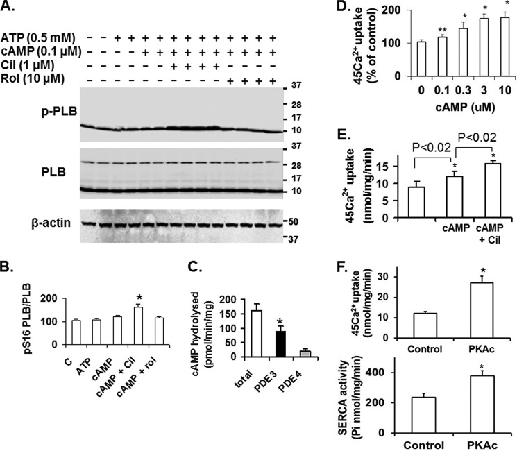
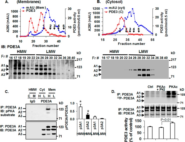
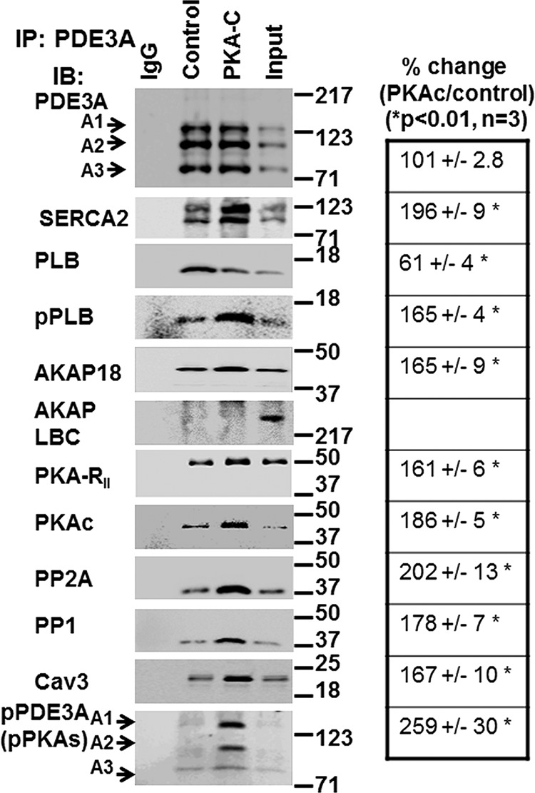
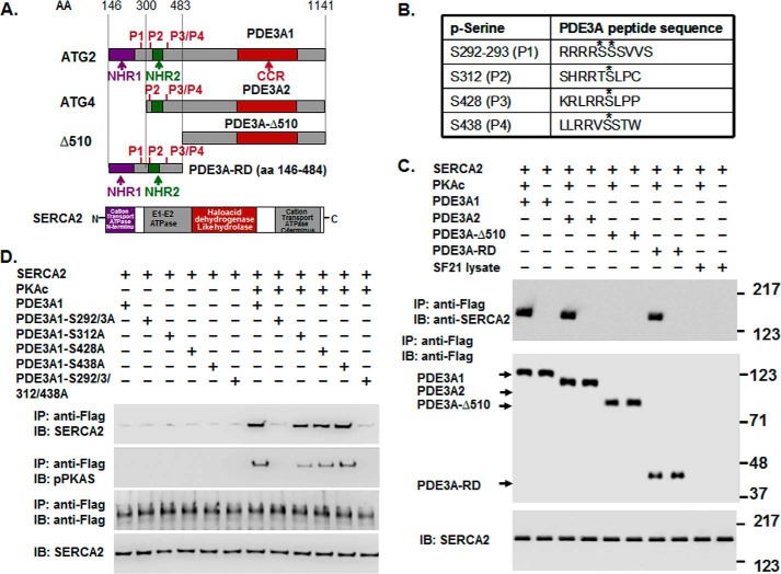
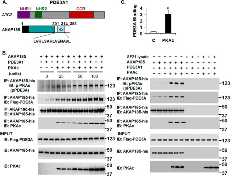
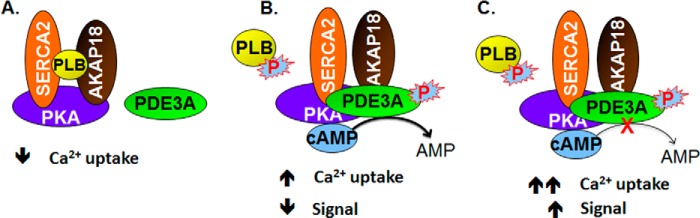
Cyclic nucleotide phosphodiesterase 3A (PDE3) regulates cAMP-mediated signaling in the heart, and PDE3 inhibitors augment contractility in patients with heart failure. Studies in mice showed that PDE3A, not PDE3B, is the subfamily responsible for these inotropic effects and that murine PDE3A1 associates with sarcoplasmic reticulum Ca(2+) ATPase 2 (SERCA2), phospholamban (PLB), and AKAP18 in a multiprotein signalosome in human sarcoplasmic reticulum (SR). Immunohistochemical staining demonstrated that PDE3A co-localizes in Z-bands of human cardiac myocytes with desmin, SERCA2, PLB, and AKAP18. In human SR fractions, cAMP increased PLB phosphorylation and SERCA2 activity; this was potentiated by PDE3 inhibition but not by PDE4 inhibition. During gel filtration chromatography of solubilized SR membranes, PDE3 activity was recovered in distinct high molecular weight (HMW) and low molecular weight (LMW) peaks. HMW peaks contained PDE3A1 and PDE3A2, whereas LMW peaks contained PDE3A1, PDE3A2, and PDE3A3. Western blotting showed that endogenous HMW PDE3A1 was the principal PKA-phosphorylated isoform. Phosphorylation of endogenous PDE3A by rPKAc increased cAMP-hydrolytic activity, correlated with shift of PDE3A from LMW to HMW peaks, and increased co-immunoprecipitation of SERCA2, cav3, PKA regulatory subunit (PKARII), PP2A, and AKAP18 with PDE3A. In experiments with recombinant proteins, phosphorylation of recombinant human PDE3A isoforms by recombinant PKA catalytic subunit increased co-immunoprecipitation with rSERCA2 and rat rAKAP18 (recombinant AKAP18). Deletion of the recombinant human PDE3A1/PDE3A2 N terminus blocked interactions with recombinant SERCA2. Serine-to-alanine substitutions identified Ser-292/Ser-293, a site unique to human PDE3A1, as the principal site regulating its interaction with SERCA2. These results indicate that phosphorylation of human PDE3A1 at a PKA site in its unique N-terminal extension promotes its incorporation into SERCA2/AKAP18 signalosomes, where it regulates a discrete cAMP pool that controls contractility by modulating phosphorylation-dependent protein-protein interactions, PLB phosphorylation, and SERCA2 activity.

Keywords: A-kinase Anchoring Protein (AKAP); Cyclic AMP (cAMP); Cyclic Nucleotide Phosphodiesterase; Immunohistochemistry; PDE3A; Phospholamban; Protein Kinase A (PKA); SERCA2; Subcellular Fractionation.


Figures

FIGURE 1.
FIGURE 1.
PDE3A, SERCA2, PLB, and AKAP18 co-localize in the Z-bands in normal human myocardium. A, cryostat sections of normal human left ventricle were prepared and, as described under “Methods,” permeabilized, blocked, and then incubated in blocking buffer with rabbit anti-PDE3A-CT, anti-desmin, anti-SERCA2, anti-PLB, anti-AKAP18, and anti-myomesin primary antibodies followed by incubation with Alexa Fluor® 488- or 594-conjugated anti-mouse or anti-rabbit secondary antibodies. Signals were detected with a Zeiss LSM510 laser scanning confocal microscope. From the left: first column, red fluorescent staining for marker proteins: desmin, SERCA2, myomesin, PLB, and AKAP18; second column, green fluorescence staining for PDE3A; third column, DAPI staining of nuclei (blue); fourth column, merged images of PDE3A, marker proteins, and DAPI indicate that PDE3A exhibits a striated pattern and co-localizes with desmin, SERCA2, PLB, and AKAP18. B, merged images from stacks of 10–15 sections (with 1-μm intervals) reveal colocalization of PDE3A with desmin, SERCA2, PLB, and AKAP18 but not with myomesin (labeling M-line Red). X-Y (center), above X-Z (top), and Y-Z (right) planes are at the indicated positions. Representative images from three independent experiments are shown.
FIGURE 2.
FIGURE 2.
cAMP, PKA, and PDE3 inhibition increase SERCA2 activity and Ca2+ uptake in human SR fractions. A, as described under “Methods,” after incubation of SR fractions (20 μg) in the absence or presence of the indicated concentrations of ATP and cAMP without or with cilostamide (Cil) or rolipram (Rol), endogenous PLB, phosphorylated PLB, and β-actin were detected after SDS-PAGE and immunoblotting. Data are representative of three experiments. In these and other Western blots PLB is predominantly monomeric, most likely due to the heating of samples before electrophoresis under reducing conditions with Laemmli SDS sample buffer. B, bar graph summarizing pSer16PLB/PLB total ratios. Cilostamide significantly enhanced the effect of 0.1 μm cAMP on phosphorylation of PLB. *, p < 0.01 versus control (n = 3 independent experiments). C, PDE activity in human cardiac SR fractions was assayed as described under “Methods” and expressed as specific activity (pmol of cAMP hydrolyzed/min/mg). Results are presented as the mean ± S.E. (n = 3 preparations). PDE3 activity was determined as the cilostamide-sensitive fraction, and PDE4 activity was determined as the rolipram-sensitive fraction. PDE3 activity was significantly higher than PDE4 activity (*, p < 0.001). D, after incubation of SR fractions without or with the indicated concentrations of cAMP (0–10 μm), 45Ca2+ uptake was measured in the presence of 0.5 μm free Ca2+ as described under “Methods.” Results are presented as % increase due to cAMP, with basal Ca2+ uptake (9.4 ± 0.8 nmol/mg/min, n = 3) taken as 100%. cAMP significantly enhanced 45Ca2+ uptake (*, p < .01; **, p < 0.04). E, after incubation of SR fractions with or without cAMP (3 μm) in the presence or absence of cilostamide (1 μm), 45Ca2+ uptake (0.5 μm free Ca2+) was assayed as described under “Methods.” Results are presented as the mean ± S.E. (n = 3). Cilostamide significantly enhanced the effect of cAMP on 45Ca2+ uptake (*, p < .02). F, Ca2+ uptake (upper panel) and SERCA activity (lower panel) were assayed in reaction mixtures containing Mg2+ and ATP in the presence or absence of rPKAc as described under “Methods.” Results are presented as nmol (Ca2+ or Pi)/min/mg (mean ± S.E.) (n = 3). rPKAc significantly enhanced 45Ca2+ uptake and SERCA2 activity (*, p < .001).
FIGURE 3.
FIGURE 3.
Analysis of PDE3A isoforms by S6 gel filtration chromatography of solubilized human myocardial membrane or cytosolic fractions. Upper panels: A and B, solubilized myocardial (myc) membranes (A) and cytosolic (cyt) fractions (Fr; B) were prepared (3 mg of protein, 1 ml) and subjected to chromatography on Superose 6 columns. Portions (10 μl) of fractions (0.5 ml) were assayed for PDE3 activity (▴) (pmol of cAMP hydrolyzed/min/0.5 ml) and protein content (milliabsorption units (mAU)) 280 nm) (●). Molecular mass standard peaks are indicated: 1, thyroglobulin (670 kDa); 2, γ-globulin (158 kDa); 3, ovalbumin (44 kDa); 4, myoglobin (17 kDa); 5, vitamin B12 (1.35 kDa). IB, immunoblot. Lower panels: A and B, portions (20 μl) of the indicated fractions were subjected to SDS/PAGE and immunoblotted with anti-PDE3A-CT antibodies. One representative experiment is shown (n = 3). C, as described under “Methods,” portions of pooled HMW (H) and LMW (L) fractions (Fig. 3, A and B) from cytosol (C) and solubilized membrane (M) fractions were cleared by incubation with 5 μg non-immune rabbit IgG and 50 μl of Protein G magnetic beads. The beads were removed by placing the tubes in a magnetic stand, and the cleared fractions were then incubated with 10 μg of non-immune IgG (IgG) or 10 μg of anti-PDE3A-CT (PDE3A) as indicated. Fractions were subjected to immunoprecipitation (IP) with 50 μl Protein G magnetic beads, which were separated from the HMW and LMW fractions as described above. The beads were washed, and proteins were eluted from the beads by boiling with Laemmli SDS sample buffer. Portions (15–20 μl) of the eluted proteins were subjected to SDS/PAGE and immunoblotted with anti-phospho-PKA-substrate (upper panel) and anti-PDE3A-CT (lower panel) antibodies. PDE3A and pPDE3A were detected in immunoprecipitates from cleared HMW and LMW fractions incubated with anti-PDE3A-CT but not non-immune IgG. Bar graph, right panel, pPDE3A1/PDE3A1 (p3A1/3A1) and pPDE3A2/PDE3A2 (p3A2/3A2) ratios in HMW and LMW peaks indicated that endogenous PDE3A1 (HMW peak) was the most highly PKA-phosphorylated isoform and demonstrated an ≈8-fold increase in phosphorylation of PDE3A1 and ∼5-fold increase in phosphorylation of PDE3A2 in HMW peaks compared with LMW peaks (*, p < 0.01). Results are representative of three individual experiments. D, as described under “Methods,” PDE3A was immunoprecipitated by incubation of cleared solubilized myocardial membranes with anti-PDE3A-CT and Protein G magnetic beads. The beads containing immunoprecipitated PDE3A were separated from the solubilized membranes, washed, and incubated as indicated without (Ctrl) or with 250 units of rPKAc or 250 units rPKAc plus 10 μm PKI (PKAc inhibitor peptide) in phosphorylation buffer containing 200 μm ATP and 5 mm MgCl2 supplemented with (upper panel) or without (lower panel) [γ-32P]ATP. Upper panel, after incubation in phosphorylation buffer containing [γ-32P]ATP, the beads containing immunoprecipitated PDE3A were separated from reaction mixtures and washed, and 32P-labeled proteins were eluted from the beads by boiling with Laemmli SDS sample buffer. Portions (15–20 μl) of the eluted proteins were subjected to SDS/PAGE. γ-32P phosphorylation of PDE3A was detected by scanning the wet gels by phosphorimaging (GE Healthcare). Phosphorylation was blocked by PKI. Middle panel, proteins on wet gels were then electrophoretically transferred to nitrocellulose membranes, and PDE3A was identified by Western blotting using anti-PDE3A-CT antibody. Lower panel, PDE3 activity was assayed in PDE3A immunoprecipitates incubated with or without rPKAc and PKI in the absence of [γ-32P]ATP. Results are expressed as pmol of cAMP hydrolyzed/min. Shown are representative data from three independent experiments. rPKAc significantly increased PDE3A activity (p < 0.01); activation was blocked by PKI (PKAc inhibitor peptide).
FIGURE 4.
FIGURE 4.
Treatment of LMW fractions of solubilized human myocardial membranes with rPKAc induces the shift of PDE3A isoforms and components of the SERCA2 regulatory signalosome into HMW fractions during S6 gel filtration chromatography. Solubilized myocardial membranes (3 mg of protein, 1 ml) were subjected to chromatography on S6 columns as described in Fig. 3A and fractionated into LMW and HMW fractions. As described under “Methods,” LMW fractions were pooled from two different experiments (cf. Fig. 3A) and concentrated via Centriprep YM-3. Upper panels, pooled and concentrated Superose 6 LMW fractions were split, incubated (1 h, 30 °C) in phosphorylation buffer with 200 μm ATP and 5 mm MgCl2 in the absence or presence of rPKAc, and re-chromatographed on Superose 6. Portions (10 μl) of fractions (0.5 ml) were assayed for PDE3 activity (▴) (pmol of cAMP hydrolyzed/min/0.5 ml) and protein content (milliabsorption units (mAU)) 280 nm) (●). Molecular mass standards: 1, thyroglobulin (670 kDa); 2, γ-globulin (158 kDa); 3, ovalbumin (44 kDa); 4, myoglobin (17 kDa); 5, vitamin B12 (1.35 kDa). Bottom panels, portions (20 μl) of the indicated fractions were subjected to SDS/PAGE and immunoblotted (IB) with the indicated antibodies. Representative results from one of two independent experiments are shown. Cav3, caveolin 3.
FIGURE 5.
FIGURE 5.
rPKAc promotes interactions of PDE3A with components of the SERCA2 regulatory signalosome. Solubilized myocardial membranes were prepared (3 mg of protein, 1 ml) and subjected to chromatography on Superose 6 columns as in Fig. 3A. Membrane LMW fractions were pooled from two different experiments (Fig. 3A) and concentrated via Centriprep YM-3. Pooled, concentrated fractions were split into three fractions and incubated in phosphorylation buffer with 200 μm ATP and 5 mm MgCl2 for 1 h at 30 °C in the absence (IgG, Control) or presence (PKA-C) of rPKAc. At the completion of these reactions, fractions were cleared with non-immune IgG and Protein G magnetic beads as described above and then incubated (overnight, 4 °C) with non-immune IgG (IgG) or anti-PDE3A-CT antibody (control, PKA-C) before incubation and immunoprecipitation (IP) with Protein G magnetic beads (1 h, 4 °C). Proteins associated with the Protein G magnetic beads were eluted by boiling in 200 μl of Laemmli SDS sample buffer. Samples (15 μl) were subjected to SDS/PAGE and immunoblotted (IB) with specific antibodies as shown. Input membrane proteins (10 μg) were also loaded on the gels as positive controls. Representative results from three independent experiments are shown. Similar amounts of PDE3A were immunoprecipitated in the control fractions and in fractions incubated with rPKAc. Band intensities of immunoprecipitated PDE3A and its interacting signaling molecules were analyzed using an LAS3000 analyzer and presented as binding percentage ratios of signaling molecules (rPKAc/control). For PDE3A, band intensities of pPDEA1/pPDE3A2/pPDE3A3 in PKAc/control percentage ratios were calculated. *, p < 0.01 versus control (n = 3 independent experiments) Cav3, caveolin 3.
FIGURE 6.
FIGURE 6.
rPKAc phosphorylates rhPDE3A and increases its interactions with rSERCA2. A, schemes representing protein domains and phosphorylation sites in FLAG-tagged-PDE3A isoforms (NHR1, transmembrane domain (obligatory membrane insertion domain); NHR2, membrane association domain; CCR, conserved C-terminal catalytic region; P1–4, predicted PKA phosphorylation sites; rhPDE3A and truncated mutants (PDE3A2, PDE3A-Δ510, PDE3A-RD (regulatory domain: aa 146–484)) and protein domains in rSERCA2 (1042 amino acids, including 3–77 cation transporter N termini; 93–341, E1-E2 ATPase; 345–724, haloacid dehydrogenase like hydrolase; 819–991, cation transporter ATPase C terminus). B, putative PKA phosphorylation sites in PDE3A designated with asterisks. C and D, purified rSERCA2 (150 ng) (Abnova) and 50 units of FLAG-tagged rhPDE3A1 or FLAG-tagged truncated mutants ((rhPDE3A2, rhPDE3A-Δ510, and rhPDE3A-RD) (C) or FLAG-tagged mutants lacking the PKA putative phosphorylation sites of rhPDE3A1 (S292A/S293A (P1), S312A (P2), S428A (P3), S438A (P4), S292A/S293A/S312A/S438A (P5)) (D) were incubated for 30 min at 30 °C in phosphorylation buffer containing 5 mm MgCl2 and 200 μm ATP in the presence or absence of rPKAc. As described under “Methods,” proteins were immunoprecipitated (IP) with anti-FLAG M2 magnetic beads. Immunoprecipitated proteins bound to the anti-FLAG-M2 magnetic beads were eluted by boiling in Laemmli SDS buffer (200 μl), and samples (15–20 μl) of eluted proteins as well as of reaction mixtures (lowest panel) were subjected to SDS-PAGE and Western immunoblotting (IB) as indicated with anti-SERCA2, anti-PKA substrate, and anti-FLAG-HRP primary antibodies and anti-mouse-HRP or anti-rabbit-HRP secondary antibodies as needed. Lowest panels, input control (SERCA2). Shown are representative blots from three independent experiments.
FIGURE 7.
FIGURE 7.
rPKAc-induced phosphorylation of rhPDE3A increases its interaction with rat rAKAP18δ. A, schemes representing hPDE3A1 protein domains (NHR1, trans-membrane domain (obligatory membrane insertion domain); NHR2, membrane association domain; CCR, conserved C-terminal catalytic region) and rAKAP18δ protein domains, with RII binding site (aa 301–314) and a unique N terminus (aa 1–26). B and C, His-tagged rAKAP18δ (100 ng) and 50 units of FLAG-tagged rhPDE3A1 were incubated for 30 min at 30 °C in phosphorylation buffer containing 200 μm ATP and 5 mm MgCl2 in the absence or presence of the indicated concentrations (units) of rPKAc (B) or in the absence or presence of 50 units of rPKAc (C). B and C, as described under “Methods,” proteins were immunoprecipitated (IP) with anti-His mAb magnetic beads, and immunoprecipitated proteins bound to the anti-His mAb magnetic beads were eluted by boiling in Laemmli SDS buffer (200 μl). Samples (15–20 μl) of eluted proteins as well as of reaction mixtures (Input, lower panels) were subjected to SDS-PAGE and Western immunoblotted (IB) with anti-FLAG-HRP, anti-AKAP18-His, anti-PKAc, and anti-phospho-PKA substrate (pPDE3A) antibodies as indicated. Similar amounts of AKAP18δ were immunoprecipitated in the control groups and in reactions incubated with rPKAc. C, as a control, rhPDE3A1 (50 units) was incubated with or without rPKAc (50 units) and with SF21 cell supernatants that contained or did not contain rAKAP18δ. The bar graph summarizes binding of rhPDE3A1-FLAG with rAKAP18δ-His in the absence (control) and presence of rPKAc. The band density of rhPDE3A1 was normalized to that of input rAKAP18δ and calculated as the amounts of rhPDE3A1 bound to rAKAP18δ in the absence (Control, C) or presence of rPKAc; there was an ∼9-fold increase in the association of rhPDE3A1 with AKAP18δ in the presence of rPKAc (*, p < 0.001). Shown are representative blots from three independent experiments.
FIGURE 8.
FIGURE 8.
Model of the regulation of SERCA2 activity by cAMP and the AKAP18 and PLB-containing signalosome. A, components of the AKAP18/SERCA2/PLB signalosome are shown. B, in the absence of cAMP, SERCA2 was inhibited by its interaction with PLB. Activation of PKA by cAMP resulted in the phosphorylation of PLB and PDE3A (and, most likely, other molecules in the signalosome). The former dissociates from SERCA2, increasing SERCA2 activity, but the integration of phosphorylated PDE3A into the signalosome limits this effect by increasing hydrolysis of cAMP. PP1 and PP2A in the signalosome would be expected to catalyze the dephosphorylation of PDE3A, PLB, and other PKA substrates and return the SERCA2 complex to its basal state. C, PDE3 inhibition potentiates the effect of cAMP on SERCA2.

References

    1. Sun B., Li H., Shakur Y., Hensley J., Hockman S., Kambayashi J., Manganiello V. C., Liu Y. (2007) Role of phosphodiesterase type 3A and 3B in regulating platelet and cardiac function using subtype-selective knockout mice. Cell. Signal. 19, 1765–1771 - PubMed
    1. Beca S., Ahmad F., Shen W., Liu J., Makary S., Polidovitch N., Sun J., Hockman S., Chung Y. W., Movsesian M., Murphy E., Manganiello V., Backx P. H. (2013) Phosphodiesterase type 3A regulates basal myocardial contractility through interacting with sarcoplasmic reticulum calcium ATPase type 2a signaling complexes in mouse heart. Circ. Res. 112, 289–297 - PMC - PubMed
    1. Lygren B., Carlson C. R., Santamaria K., Lissandron V., McSorley T., Litzenberg J., Lorenz D., Wiesner B., Rosenthal W., Zaccolo M., Taskén K., Klussmann E. (2007) AKAP complex regulates Ca2+ re-uptake into heart sarcoplasmic reticulum. Embo. Rep. 8, 1061–1067 - PMC - PubMed
    1. Stefan E., Wiesner B., Baillie G. S., Mollajew R., Henn V., Lorenz D., Furkert J., Santamaria K., Nedvetsky P., Hundsrucker C., Beyermann M., Krause E., Pohl P., Gall I., MacIntyre A. N., Bachmann S., Houslay M. D., Rosenthal W., Klussmann E. (2007) Compartmentalization of cAMP-dependent signaling by phosphodiesterase-4D is involved in the regulation of vasopressin-mediated water reabsorption in renal principal cells. J. Am. Soc. Nephrol. 18, 199–212 - PubMed
    1. Ahmad F., Lindh R., Tang Y., Weston M., Degerman E., Manganiello V. C. (2007) Insulin-induced formation of macromolecular complexes involved in activation of cyclic nucleotide phosphodiesterase 3B (PDE3B) and its interaction with PKB. Biochem. J. 404, 257–268 - PMC - PubMed

Publication types

MeSH terms

Substances