. 2015 Mar 5;125(10):1633-42.
doi: 10.1182/blood-2014-08-597419. Epub 2015 Jan 15.

Defective PDI release from platelets and endothelial cells impairs thrombus formation in Hermansky-Pudlak syndrome

Affiliations

Defective PDI release from platelets and endothelial cells impairs thrombus formation in Hermansky-Pudlak syndrome

Anish Sharda et al. Blood. .

Abstract

Protein disulfide isomerase (PDI), secreted from platelets and endothelial cells after injury, is required for thrombus formation. The effect of platelet and endothelial cell granule contents on PDI-mediated thrombus formation was studied by intravital microscopy using a mouse model of Hermansky-Pudlak syndrome in which platelet dense granules are absent. Platelet deposition and fibrin generation were nearly absent, and extracellular PDI was significantly reduced in HPS6(-/-) mice after vascular injury. HPS6(-/-) platelets displayed impaired PDI secretion and impaired exocytosis of α granules, lysosomes, and T granules due to decreased sensitivity to thrombin, but these defects could be corrected by addition of subthreshold amounts of adenosine 5'-diphosphate (ADP). Human Hermansky-Pudlak syndrome platelets demonstrated similar characteristics. Infusion of wild-type platelets rescued thrombus formation in HPS6(-/-) mice. Human umbilical vein endothelial cells in which the HPS6 gene was silenced displayed impaired PDI secretion and exocytosis of Weibel-Palade bodies. Defective thrombus formation in Hermansky-Pudlak syndrome, associated with impaired exocytosis of residual granules in endothelial cells and platelets, the latter due to deficiency of ADP, is characterized by a defect in T granule secretion, a deficiency in extracellular PDI secretion, and impaired fibrin generation and platelet aggregation. Hermansky-Pudlak syndrome is an example of a hereditary disease whereby impaired PDI secretion contributes to a bleeding phenotype.


Figures

Figure 1
Figure 1
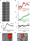
Platelet thrombus formation, fibrin generation, and PDI secretion after laser-induced arteriolar wall injury in HPS6−/− and WT mice. Platelet-specific anti-CD42b antibody and fibrin-specific mouse anti-human fibrin monoclonal antibody were infused into mice, and the cremaster arteriole subjected to laser injury and the induction of thrombus formation. (A) Representative images of the fluorescence associated with fibrin (green) and platelets (red) over 180 seconds after laser-induced vessel wall injury. (B) Median integrated platelet fluorescence intensity vs time in WT mice (dark red) and HPS6−/− mice (red). (C) Median integrated fibrin fluorescence intensity vs time in WT mice (dark green) and HPS6−/− mice (green). Data are from 30 thrombi in 3 mice for HPS6−/− and WT mice. In panels A-C, platelet-specific anti-CD42b antibody and nonblocking polyclonal anti-PDI antibody were infused into mice to detect platelets and PDI during thrombus formation. (D) Representative images of fluorescence associated with PDI (green) and platelets (red) over 180 seconds of thrombus formation after laser-induced vessel wall injury in WT and HPS6−/− mice. (E) Median integrated platelet fluorescence intensity during thrombus formation in WT mice (dark red) and HPS6−/− mice (red). (F) Median integrated PDI fluorescence intensity during thrombus formation in WT mice (dark green) and HPS6−/− mice (green). F, fluorescence intensity.
Figure 2
Figure 2
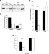
Localization of PDI and granule release in WT and HPS6−/− platelets. (A) Localization of PDI in platelet granules. (a) Unstimulated platelets isolated from WT and HPS6−/− mice were sedimented by centrifugation. The pellet was solubilized with SDS lysis buffer to generate the lysate. Lysate of intact resting platelets from WT and HPS6−/− mice was subjected to SDS-PAGE and blotted with anti-PDI antibodies DL-11. (b) Washed WT mouse resting platelets were fixed, frozen, and sectioned before mounting on Formvar carbon-coated copper grids. Ultrathin platelet sections were probed for PDI, and bound antibody labeled with Protein A-gold. Samples were examined by transmission electron microscopy and reveal distribution of PDI in platelet T granules. Bar represents 100 nm. (c) As per panel Ab, but HPS6−/− mouse platelets were examined. (B) Decreased thrombin sensitivity of granule exocytosis in HPS6−/− platelets. Comparing WT platelets and HPS6−/− platelets, thrombin-induced granule exocytosis was studied in vitro to characterize α granule, T granule, and lysosomes. Mouse platelets in HTG buffer (250 × 105 platelets per 100 μL of 5 mM d-glucose, 134 mM sodium chloride, 0.34 mM disodium phosphate, 2.9 mM potassium chloride, 12 mM sodium bicarbonate, 20 mM N-2-hydroxyethylpiperazine-N′-2-ethanesulfonic acid, and 1 mM magnesium chloride, pH 7.3) were incubated with mouse α-thrombin for 10 minutes at room temperature. Twenty microliters of thrombin-stimulated or resting platelets were incubated with fluorescein isothiocyanate–conjugated P-selectin, TLR9, or Alexa Fluor 488–labeled LAMP1 antibodies for 15 minutes. Surface expression of platelet granule markers was measured using CellQuest on a FACSCalibur flow cytometer (Becton Dickinson). Data are expressed as percent maximal release. Immunoflow cytometry of P-selectin monitored α granule exocytosis, whereas α granule content release, monitored by PF-4 secretion, was measured by enzyme-linked immunosorbent assay. T granule exocytosis was monitored by surface exposure of TLR9 using flow cytometry and lysosome exocytosis by the release of LAMP1. For PF-4 and PDI, hirudin (1 U/mL) was added to quench thrombin activity, then platelets were sedimented by centrifugation, the supernatant was collected and ultracentrifuged at 71 000g for 30 minutes, and the releasate was assayed. Platelets from WT or HPS6−/− mice were activated with varying amounts of thrombin, and the markers for granule exocytosis were measured. Thrombin agonist concentrations were 0.007, 0.036, 0.072, 0.36, 0.72, 3.6, and 7.2 nM; the average of 5 measurements defines each point ± SD. (a) P-selectin (α granules); (b) PF-4 (α granules); (c) TLR9 (T granules); (d) LAMP 1 (lysosomes); **P < .01, ***P < .001. (e) Band densities of PDI antigen in releasates of thrombin-stimulated WT and HPS6−/− platelets detected by SDS-PAGE, followed by immunoblotting with anti-PDI antibodies (DL-11; 1 μg/mL). Data represent mean ± SD (n = 2; **P < .01). (f) Thiol isomerase secretion after platelet activation with 0.72 or 7.2 nM thrombin. Thiol isomerase activity was monitored by the reduction of a di-E-GSSG as a substrate. The increase in fluorescence was measured at excitation/emission of 525/540 nm for 20 minutes at 25°C. **P < .01. WT platelets, closed bars; HPS6−/− platelets, open bars. (C) Agonist-induced adenosine triphosphate release as a marker of dense granule release in WT and HPS6−/− platelets as measured by luminometry. WT and HPS6−/− platelets were activated with SFLLRN (SF; 252 μM), collagen (Col; 19 μg/mL), or buffer control (Control), and the kinetics of release of adenosine triphosphate was monitored as a function of time (0, 5, 10, 15, 20, 25, and 30 seconds, left to right) by luminometry. (D) Decreased collagen sensitivity of granule exocytosis from HPS6−/− platelets. Band densities of PDI antigen in releasates of collagen-stimulated (0.1-5 μg/mL of type 1 equine collagen; 10 minutes) WT and HPS6−/− platelets detected by SDS-PAGE, followed by immunoblotting with anti-PDI antibodies (DL-11; 1 μg/mL) (n = 3; mean ± SD; **P = .001, *P = .01). WT platelets, closed bars; HPS6−/− platelets, open bars. SD, standard deviation; SDS-PAGE, sodium dodecyl sulfate–polyacrylamide gel electrophoresis.
Figure 3
Figure 3
Rescue of thrombin-induced PDI release from HPS6−/− platelets using substimulatory ADP, and reduction of PDI release from WT platelets with apyrase. PDI antigen in releasate of thrombin-stimulated (0.36 nM) WT and HPS6−/− platelets with and without substimulatory concentrations of ADP (1 μM) or with and without apyrase (Apy; 0.1 U/mL). Samples were obtained 10 minutes after thrombin activation. (A) PDI antigen in releasates of thrombin-stimulated WT and HPS6−/− platelets were detected by SDS-PAGE, followed by immunoblotting with anti-PDI antibodies (DL-11; 1 mg/mL). (B) Bar graph comparing PDI release in WT and HPS6−/− platelets in the presence (+) or absence (-) of ADP. Data represent mean ± SD; n = 2; **P < .01. (C) Bar graph comparing PDI release in WT and HPS6−/− platelets in the presence (+) or absence (-) of apyrase. Data represent mean ± SD (n = 2; *P < .01). (D) Effect of high levels of ADP on HPS6−/− release of PDI. WT and HPS6−/− platelets were incubated with 10 μM (-) or 100 μM (+) of ADP, and releasate was collected and resolved on SDS-PAGE. PDI was quantitated by immunoblotting with rabbit polyclonal anti-PDI antibody (n = 2; mean ± SD; **P = .02). WT, closed bars; HPS6−/−, open bars.
Figure 4
Figure 4
Rescue of thrombus formation in HPS6−/− mice by infusion of WT platelets. Washed platelets isolated from WT mice were infused into HPS6−/− mice to a final approximate concentration of 10% to 20% of the total mouse platelet count. Platelet-specific anti-CD42b antibody and fibrin-specific mouse anti-human fibrin monoclonal antibody were infused to detect platelets and fibrin during in vivo thrombus formation initiated by laser-induced injury. (A) Representative images of the fluorescence associated with fibrin (green) and platelets (red) over 180 seconds after laser-induced vessel wall injury in an HPS6−/− mouse (left) and an HPS6−/− mouse treated with WT mouse platelets (right). (B) Median integrated platelet fluorescence intensity as a function of time in the absence (red) and presence (dark red) of WT platelets. (C) Median integrated fibrin fluorescence intensity as a function of time in the absence (green) and presence (dark green) of WT platelets. Under identical experimental conditions, a nonblocking anti-PDI antibody was employed instead of the fibrin-specific monoclonal antibody to visualize PDI. (D) Median integrated platelet fluorescence intensity as a function of time in the absence (gray) and presence (black) of infused WT platelets. (E) Median integrated PDI fluorescence intensity as a function of time in the absence (gray) and presence (black) of infused WT platelets. Data in panels B-E are from 30 thrombi in 3 mice for each condition. (F) Distribution of calcein-labeled WT donor platelets in a developing thrombus following their infusion into an HPS6−/− mouse. (G) Distribution of calcein-labeled WT donor platelets in a developing thrombus following their infusion into a WT mouse. Donor WT platelets, green; platelets, red; merge, yellow.
Figure 5
Figure 5
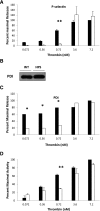
Hermansky-Pudlak syndrome in humans: impaired α granules and PDI secretion in a patient with Hermansky-Pudlak syndrome. Comparing normal human platelets and human Hermansky-Pudlak syndrome platelets, thrombin-induced granule exocytosis was studied in vitro to characterize α granule release, thiol isomerase secretion, and PDI antigen secretion. Platelet activation was performed with indicated amounts of thrombin. (A) P-selectin (α granules) expression by flow cytometry in platelets from a Hermansky-Pudlak syndrome patient and from a normal subject. (B) PDI antigen in lysate of resting normal and Hermansky-Pudlak syndrome platelets. (C) PDI antigen secretion monitored by western blot analysis of the releasate of thrombin-activated normal and Hermansky-Pudlak syndrome platelets. (D) Thiol isomerase activity monitored by the reduction of a di-E-GSSG as a substrate. Normal platelets, closed bars; Hermansky-Pudlak syndrome platelets, open bars. N = 2 experiments; mean ± SD; *P = .02, **P < .01.
Figure 6
Figure 6
HPS6 knockdown in HUVECs treated with HPS6 siRNA. HPS6 in HUVECs 48 and 72 hours posttransfection with HPS6 or control siRNA were detected by SDS-PAGE of cell lysates followed by immunoblotting with polyclonal anti-HPS6 antibodies (HPS6d; 2.5 μg/mL). (A) Band densities of HPS6 at 48 and 72 hours posttransfection with control siRNA (closed bars) or HPS6 siRNA (open bars). Control is shown as 100%. Data represent mean ± SD (n = 3; ***P < .001). (B) Immunoblot of HPS6 (50 μL of lysate from confluent monolayer, ∼0.25 × 10⁶ cells) 72 hours posttransfection with control siRNA (lanes 1, 2, and 3) or HPS6 siRNA (lanes 4, 5, and 6). The membranes were immunoblotted for GAPDH (polyclonal anti-GAPDH, 1 μg/mL) as a loading control. (C) HPS6 knockdown does not affect PDI storage in HUVECs. PDI and HPS6 in HUVECs 72 hours posttransfection with HPS6 or control siRNA were detected by SDS-PAGE of cell lysates, followed by immunoblotting with polyclonal anti-PDI antibody (DL-11; 1 μg/mL) and polyclonal anti-HPS6 antibody (HPS6d; 2.5 μg/mL), respectively. Immunoblot of PDI and HPS6 (25 μL of lysate from confluent monolayer, ∼0.25 × 10⁶ cells) 72 hours posttransfection. HUVEC lysate from cells treated with control siRNA (lanes 1, 2, and 3) or HPS6 siRNA (lanes 4, 5, and 6). Recombinant PDI (15 ng) was run as positive control. GAPDH, glyceraldehyde-3-phosphate dehydrogenase.
Figure 7
Figure 7
Thrombin-induced PDI and VWF release from HUVECs treated with HPS6 siRNA. Thrombin-induced PDI release and VWF release from HUVECs 72 hours posttransfection with HPS6 or control siRNA were detected by SDS-PAGE, followed by immunoblotting with anti-PDI antibodies (DL-11; 1 μg/mL) and anti-VWF antibodies (rabbit polyclonal; 1 μg/mL), respectively. Band densities of released PDI from HUVECs (A) or VWF released from HUVECs (B) over 30 minutes after thrombin stimulation (1 NIH U/mL) 72 hours posttransfection with control siRNA (closed bars) or HPS6 siRNA (open bars). Control is shown as 100%. Data represent mean ± SD (n = 3; ***P < .001, **P < .01). (C) When eptifibatide (10 μg/g mouse) was infused, platelet accumulation was prevented in WT mice (black) and in HPS6−/− mice (gray). (D) Eptifibatide did not blunt the median integrated PDI fluorescence intensity during thrombus formation in WT mice (black) but did so in HPS6−/− mice (gray). These results are consistent with defective PDI release from the endothelium in HPS6−/− mice. Data in panels C and D are from 30 thrombi in 3 mice for HPS6−/− and for WT mice.

Comment in

References

    1. Logan LJ, Rapaport SI, Maher I. Albinism and abnormal platelet function. N Engl J Med. 1971;284(24):1340–1345. - PubMed
    1. Hardisty RM, Mills DC, Ketsa-Ard K. The platelet defect associated with albumism. Br J Haematol. 1972;23(6):679–692. - PubMed
    1. Zhang Q, Zhao B, Li W, et al. Ru2 and Ru encode mouse orthologs of the genes mutated in human Hermansky-Pudlak syndrome types 5 and 6. Nat Genet. 2003;33(2):145–153. - PubMed
    1. Gautam R, Chintala S, Li W, et al. The Hermansky-Pudlak syndrome 3 (cocoa) protein is a component of the biogenesis of lysosome-related organelles complex-2 (BLOC-2). J Biol Chem. 2004;279(13):12935–12942. - PubMed
    1. Di Pietro SM, Falcón-Pérez JM, Dell’Angelica EC. Characterization of BLOC-2, a complex containing the Hermansky-Pudlak syndrome proteins HPS3, HPS5 and HPS6. Traffic. 2004;5(4):276–283. - PubMed

Publication types

MeSH terms