

Accurate definition and management of idiopathic sclerosing encapsulating peritonitis
- PMID: 25593498
- PMCID: PMC4292304
- DOI: 10.3748/wjg.v21.i2.675
Accurate definition and management of idiopathic sclerosing encapsulating peritonitis
Abstract
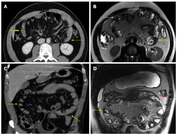
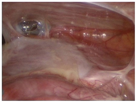
Aim: To review the literature on idiopathic sclerosing encapsulating peritonitis (SEP), also known as abdominal cocoon syndrome.
Methods: The PubMed, MEDLINE, Google Scholar, and Google databases were searched using specific key words to identify articles related to idiopathic SEP. These key words were "sclerosing encapsulating peritonitis," "idiopathic sclerosing encapsulating peritonitis," "abdominal cocoon," and "abdominal cocoon syndrome." The search included letters to the editor, case reports, review articles, original articles, and meeting presentations published in the English-language literature from January 2000 to May 2014. Articles or abstracts containing adequate information about age, sex, symptom duration, initial diagnosis, radiological tools, and surgical approaches were included in the study. Papers with missing or inadequate data were excluded.
Results: The literature search yielded 73 articles on idiopathic (primary) SEP published in 23 countries. The four countries that published the greatest number of articles were India (n = 21), Turkey (n = 14), China (n = 8) and Nigeria (n = 3). The four countries that reported the greatest number of cases were China (n = 104; 53.88%), India (n = 35; 18.13%), Turkey (n = 17; 8.80%) and Nigeria (n = 5; 2.59%). The present study included 193 patients. Data on age could be obtained for 184 patients (range: 7-87 years; mean ± SD, 34.7 ± 19.2 years), but were unavailable for nine patients. Of the 184 patients, 122 were male and 62 were female; sex data could not be accessed in the remaining nine patients. Of the 149 patients whose preoperative diagnosis information could be obtained, 65 (43.6%) underwent operations for abdominal cocoon, while the majority of the remaining patients underwent operations for a presumed diagnosis of intestinal obstruction and/or abdominal mass. Management information could be retrieved for 115 patients. Of these, 68 underwent excision + adhesiolysis (one laparoscopic); 24 underwent prophylactic appendectomy in addition to excision + adhesiolysis. Twenty patients underwent various resection and repair techniques along with excision + adhesiolysis. The remaining three patients were managed with antituberculosis therapy (n = 2) and immunosuppressive therapy (n = 1).
Conclusion: Idiopathic SEP is a rare disorder characterized by frequently recurring bouts of intestinal obstruction. Surgical therapy is the gold standard management strategy.
Keywords: Abdominal cocoon syndrome; Idiopathic; Intestinal obstruction; Primary; Sclerosisis encapsulation peritonitis.
Figures
References
-
- Li N, Zhu W, Li Y, Gong J, Gu L, Li M, Cao L, Li J. Surgical treatment and perioperative management of idiopathic abdominal cocoon: single-center review of 65 cases. World J Surg. 2014;38:1860–1867. - PubMed
-
- Jovani M, Baticci F, Bonifacio C, Omodei PD, Malesci A. Abdominal cocoon or idiopathic encapsulating peritoneal sclerosis: magnetic resonance imaging. Dig Liver Dis. 2014;46:192–193. - PubMed
Publication types
MeSH terms
LinkOut - more resources
Full Text Sources
Other Literature Sources
