Synchronic nasopharyngeal and intraparotid warthin tumors: A case report and literature review
- PMID: 25593670
- PMCID: PMC4282915
- DOI: 10.4317/jced.51441
Synchronic nasopharyngeal and intraparotid warthin tumors: A case report and literature review
Abstract
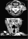
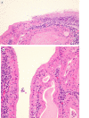
Warthin tumor is the second most frequent benign salivary gland tumor after pleomorphic adenoma; it occurs almost exclusively in the parotid gland and peri-parotideal lymph nodes, although it may rarely present in other locations. It may be multicentric and bilateral in a small percentage of cases. Nasopharyngeal Warthin tumor is very rare, and the presence of a synchronic WT involving nasopharynx and parotid is an exceptional event, as it has been described only twice in the literature. In this article we report an additional case of a synchronic Warthin tumor and review the related literature. Key words:Warthin tumor, synchronic WT, multicéntrico, nasopharynx.
Conflict of interest statement
Figures


Similar articles
-
Synchronous parotid and nasopharyngeal Warthin tumor.Head Neck. 2016 Mar;38(3):E71-2. doi: 10.1002/hed.24180. Epub 2015 Aug 28. Head Neck. 2016. PMID: 26315140
-
Low Molecular Weight Cytokeratin Immunohistochemistry Reveals That Most Salivary Gland Warthin Tumors and Lymphadenomas Arise in Intraparotid Lymph Nodes.Head Neck Pathol. 2021 Jun;15(2):438-442. doi: 10.1007/s12105-020-01215-2. Epub 2020 Aug 31. Head Neck Pathol. 2021. PMID: 32865726 Free PMC article.
-
Intraparotid classical and nodular lymphocyte-predominant Hodgkin lymphoma: pattern analysis with emphasis on associated lymphadenoma-like proliferations.Am J Surg Pathol. 2015 Sep;39(9):1206-12. doi: 10.1097/PAS.0000000000000440. Am J Surg Pathol. 2015. PMID: 25929348
-
Warthin tumor arising from the minor salivary gland.J Craniofac Surg. 2012 Sep;23(5):e374-6. doi: 10.1097/SCS.0b013e318254359f. J Craniofac Surg. 2012. PMID: 22976673 Review.
-
Presentation of Chronic Lymphocytic Leukemia/Small Lymphocytic Lymphoma in a Warthin Tumor: Case Report and Literature Review.Int J Surg Pathol. 2018 May;26(3):256-260. doi: 10.1177/1066896917734371. Epub 2017 Oct 4. Int J Surg Pathol. 2018. PMID: 28978260 Review.
Cited by
-
Oncocytic Cysts of the Nasopharynx: A Case Report.Allergy Rhinol (Providence). 2020 Sep 6;11:2152656720956594. doi: 10.1177/2152656720956594. eCollection 2020 Jan-Dec. Allergy Rhinol (Providence). 2020. PMID: 32953230 Free PMC article.
-
Synchronous parotid and nasopharyngeal Warthin's tumor: case report and literature review.Braz J Otorhinolaryngol. 2020 Dec;86 Suppl 1(Suppl 1):44-47. doi: 10.1016/j.bjorl.2017.05.009. Epub 2017 Jun 27. Braz J Otorhinolaryngol. 2020. PMID: 28711460 Free PMC article. Review. No abstract available.
References
-
- Hildebrand O. Uber angerborene epitheliale cysten und fisten des halses. Arch Klin Chir. 1895;49:167–206.
-
- Albrecht D, Arzt L. Beitrage zur frague der gewebsverrung.papillare cystadenoma in lymphdrusen. Frankfurt Z Pathol. 1910;4:47–69.
-
- Warthin AS. Papillary cystadenoma lymphomatosum. A rare teratoid of the parotid region. J Cancer Res. 1929;13:116–25.
-
- Maiorano L, Lo ML, Favia G, Piattelli A. Warthin's Tumour : a study of 78 cases with emphasis on bilaterality, multifocality and association with other malignancies. Oral Oncol. 2002;38:35–40. - PubMed
-
- Hart MN, Andrews JL. Papillary cystadenoma lymphomatosum arising in the cavity oral. Oral Surg. 1968;26:588–91. - PubMed
Publication types
LinkOut - more resources
Full Text Sources
Other Literature Sources
